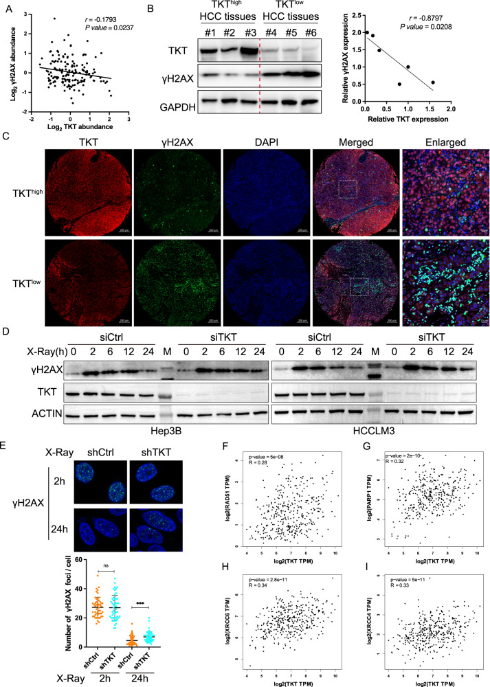
Fig. 1. TKT is involved in DSB repair in HCC.
A The correlation analysis between TKT and γH2AX expression using the published proteomic and phosphoproteomics dataset of 159 HCC patients from Fudan University. B The expression levels of γH2AX in TKT high and low tissues from 6 patients with HCC were compared using WB. C Representative MIF staining micrographs showing TKT (low or high) and γH2AX expression in an HCC microarray. Scale bar, 200 μm (TKT, γH2AX, DAPI and Merged) and 20 μm (Enlarged). D The Hep3B (Left) and HCCLM3 (Right) cells were collected without treatment, or at 2 h, 6 h, 12 h and 24 h post 4 Gy X-Ray treatment with siCtrl or siTKT transfection. Cell lysates were immunoblotted with the indicated antibodies. M, protein ladder. E Representative fluorescence images (green, γH2AX; blue, DAPI) and quantification of γH2AX immunostaining in cells at the indicated time after X-Ray treatment with or without TKT depletion. The correlation analysis between TKT and RAD51 (F), PARP1 (G), XRCC6 (H), XRCC4 (I) expression in HCC tissues by Spearman correlation analysis from TCGA database. Adapted from GEPIA: http://gepia.cancer-pku.cn/. TPM, transcripts per million.

