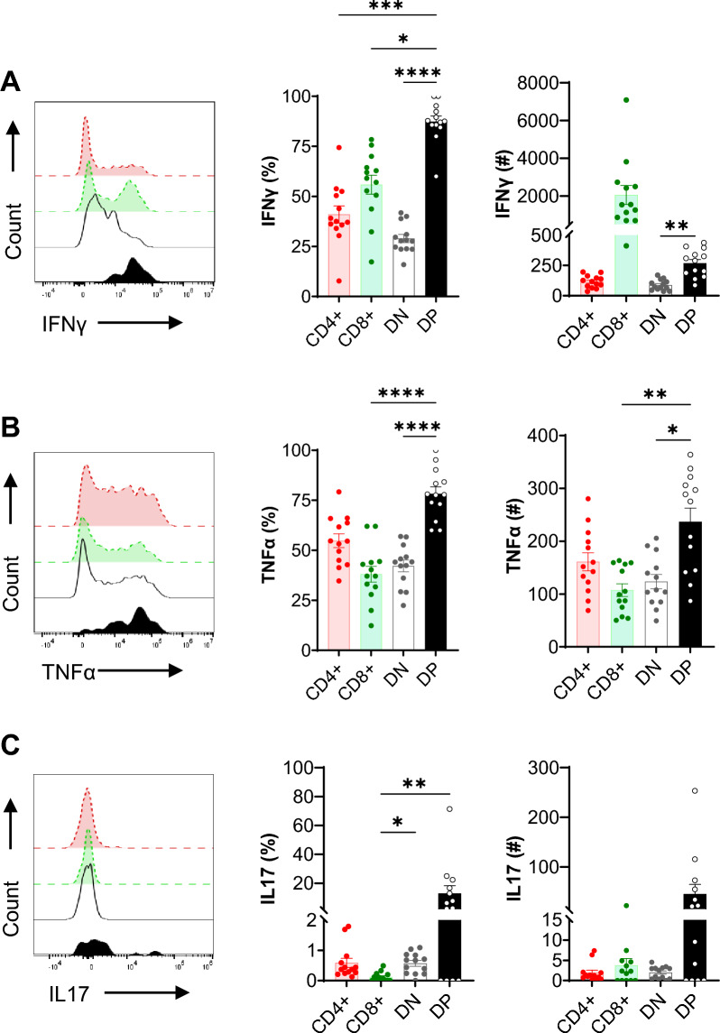
Figure 4.
Kidney DPT cells produce proinflammatory cytokines at baseline. A total of 1 × 10⁶ KMNCs were incubated with T cell activation cocktail containing PMA/Ionomycin/Brefeldin A for 4 h at 37 °C. Cells were then stained with Zombie NIR followed by surface markers (CD45, TCR, CD4, CD8) and intracellular cytokine (IFNγ, TNFα and IL-17) antibodies. Percentage (left graph) and absolute numbers (right graph) of (A) IFNγ, (B) TNFα and (C) IL-17 expressing DPT cells in normal mouse kidney (n = 13). Data are expressed as mean ± sem and compared by non-parametric Kruskal–Wallis test followed by Dunn’s post-hoc analysis. * = P ≤ 0.05, ** = P ≤ 0.01, *** = P ≤ 0.001, **** = P ≤ 0.0001.

