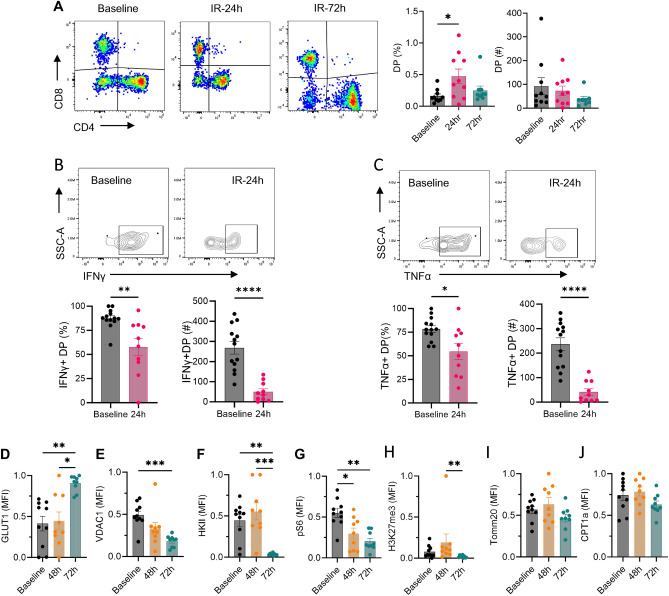
Figure 7.
Ischemia reperfusion injury increases kidney DPT cell percentage and alters their cytokine and metabolic profile. (A) Percentage (left graph) and absolute numbers (right graph) of DPT cells in WT mice subjected to 27 min bilateral ischemia reperfusion injury and euthanized either after 24 h (n = 10) or 72 h (n = 9). Mice were euthanized under the effect of Ketamine/xylazine as described in the methods section. Baseline levels of DPT cells were quantified from normal non-surgery mouse kidneys (n = 10). (B and C) KMNCs from 24 h post-IR mice (n = 10) and control baseline (n = 13) mice were isolated and assessed for cytokine expression. The percentage (left panel) and absolute number (right panel) of IFNγ and TNFα positive DPT cells decreased after IR injury. (D–J) Separate group of mice were used to study effect of IR injury on metabolic markers and euthanized at either 48 h (n = 9) or 72 h post-IR (n = 9). IR injury increased GLUT1 expression but VDAC1, HKII, pS6 and H3K27me3 were reduced compared to baseline. Data are expressed as mean ± sem and compared by non-parametric Kruskal–Wallis test followed by Dunn’s post-hoc analysis. * = P ≤ 0.05, ** = P ≤ 0.01, *** = P ≤ 0.001, **** = P ≤ 0.0001.

