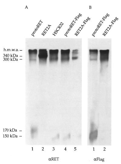
FIG. 4.
Dimer formation of the different RET mutants. (A) Equal amounts of proteins from Cos cells transfected with the protoRET, RET2A, HSCR32, protoRET-Flag, and RET2A-Flag constructs were fractionated on nonreducing SDS–7% polyacrylamide gel, transferred, and stained with anti-RET (αRET) antibodies. (B) Equivalent protein extracts from cells transfected with protoRET-Flag and RET2A-Flag constructs were processed similarly and stained with anti-Flag (αFlag) antibodies. The migration of the monomers, dimers, and high-molecular-mass aggregates (h.m.w.a.) is indicated on the left. The sizes of the dimers were determined on the basis of the migration of size markers of known molecular weights.

