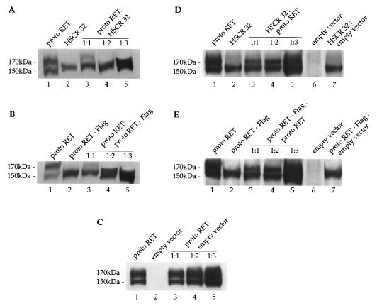
FIG. 7.
HSCR32 and protoRET-Flag mutants exert a dominant negative effect over protoRET. (A) Cos cells were cotransfected with 5 μg of protoRET and 5, 10, and 15 μg of protoRET-Flag expression vectors. Parallel transfections were carried out with 5 μg of protoRET and protoRET-Flag expression vectors alone. A Western blot analysis was performed with increasing amounts of proteins from the transfected cells. Lane 1, protoRET (50 μg); lane 2, HSCR32 (50 μg); lanes 3 to 5, protoRET and HSCR32 at ratios of 1:1 (100 μg) (lane 3), 1:2 (150 μg) (lane 4), and 1:3 (200 μg) (lane 5). After the immunoblotting, the RET species were stained with anti-RET antibodies. The migration of the 170- and 150-kDa RET monomers is indicated. (B) Western blot analysis performed as described for panel A on different amounts of protein extracts from cells individually transfected with protoRET and protoRET-Flag expression plasmids (lanes 1 and 2) or cotransfected at three molar ratios (lanes 3 to 5). The migration of the 170- and 150-kDa RET monomers is indicated. (C) Western blot analysis performed on protein extracts from cells individually transfected with protoRET or an empty vector (lanes 1 and 2) or cotransfected in three molar ratios (lanes 3 to 5). The RET species were identified by immunoblotting with anti-RET antibodies. (D) Western blot analysis performed as described for panel A on different amounts of proteins from cells transfected with protoRET and HSCR32 alone (lanes 1 and 2) or with a combination of a fixed amount of HSCR32 and three ratios of protoRET expression vector (lanes 3 to 5). In lanes 6 and 7 extracts from cells transfected with an empty vector alone or cotransfected with HSCR32 and an empty vector at a 1:3 ratio are shown. The RET species were identified with anti-RET antibodies. (E) Western blot analysis of protein extracts from cells transfected with protoRET and protoRET-Flag expression vectors alone (lanes 1 and 2) or cotransfected with a fixed amount of protoRET-Flag and three proportions of protoRET (lanes 3 to 5). Extracts from cells transfected with an empty vector or with protoRET-Flag and an empty vector at a 1:3 ratio were loaded in lanes 6 and 7. The RET species were identified with anti-RET antibodies.

