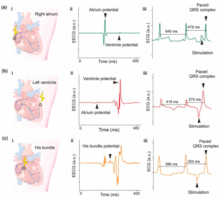
Figure 18.
Self-powered cardiac pacing in vivo. (i) Schematic diagram, (ii) epicardium electrocardiogram (EECG) of pacing sites, and (iii) typical valid pacing ECG at (a) right atrium, (b) left ventricle, and (c) His bundle. Reprinted with permission from [152]. Copyright 2023 Elsevier Ltd.

