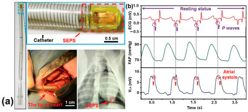
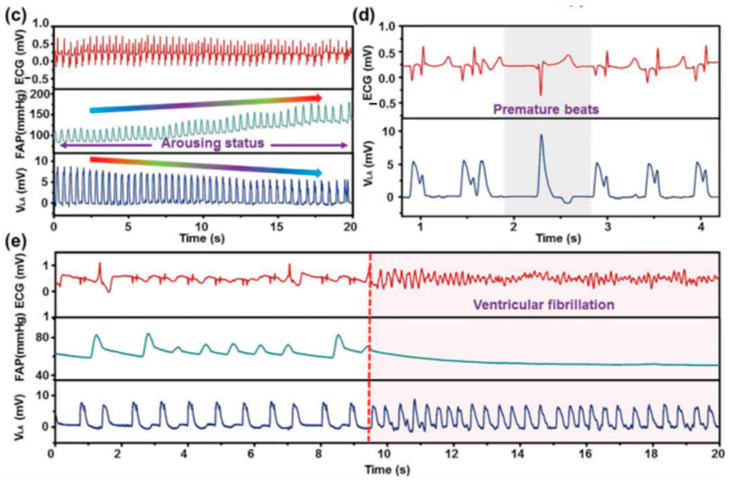
Figure 35.
(a) Photograph of minimally invasive surgery with a DR image of the heart implanted with a device by integration with a surgical delivery system. (b) Detailed inspection into the corresponding relationship between waveforms of ECG and the SEPS outputs. (c) The comparison among signals of ECG, FAP, and SEPS during the reinforcing process of cardiac function. (d) Ectopic R waves in representative ECG indicating ventricular premature contraction corresponded to an enhanced waveform of the device. (e) Disorganized waveforms of SEPS signals with quickened frequency were observed when ventricular fibrillation occurred. Reprinted with permission from [166]. Copyright 2023 Wiley-VCH.


