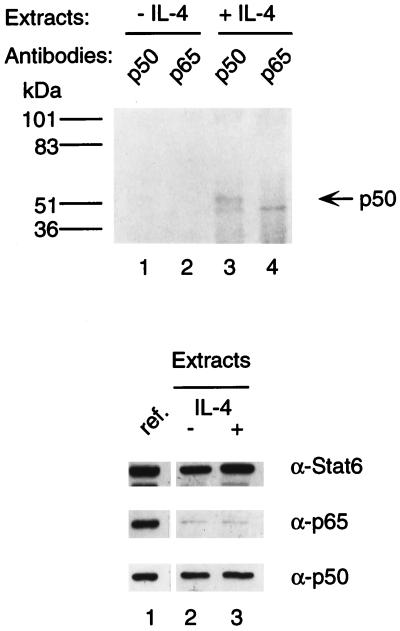
FIG. 3.
Interaction of Stat6 and NF-κB p50 in vivo. I.29μ B cells were labeled with [35S]methionine and [35S]cysteine for 3 h. Cells were then treated with LPS for 2 h, followed by stimulation with (lanes 3 and 4) or without (lanes 1 and 2) IL-4 for 30 min. Nuclear extracts were first immunoprecipitated with anti-Stat6 antibodies. Immunoprecipitates were dissolved, and aliquots were reprecipitated with rabbit antiserum against NF-κB p50 or p65. The precipitated proteins were analyzed by SDS–7.5% PAGE and fluorography. The positions of size markers were as indicated (in kilodaltons). Bottom panels: The contents of Stat6 and NF-κB proteins in the nuclear extracts (5 μg each) were analyzed by SDS–10% PAGE, followed by Western blotting with anti-Stat6, anti-p50, or anti-p65 antibody. The samples were analyzed on the same blot and autoradiographed for the same time as the reference samples (lane 1 of each panel) of nuclear extracts (5 μg each) from HEK 293 cells cotransfected with equal amounts of Stat6, NF-κB p50, and NF-κB p65 expression plasmids.

