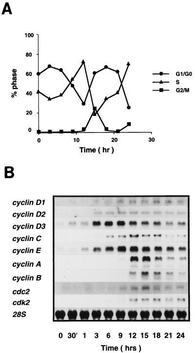
FIG. 1.
(A) Cell cycle analysis of BAF-B03 cells following IL-3 stimulation. Cells were synchronized by growth factor starvation for 15 h and restimulated with IL-3. Samples were harvested at various times after stimulation, stained with propidium iodide, and analyzed by flow cytometry as described in Materials and Methods. The calculated percentages of cells at each phase are plotted. (B) Differential expression of cyclin and cdc2 family kinase genes in BAF-B03-derived transformants stimulated with IL-3. Stimulated cells were harvested at various times as indicated, and total RNA extracted from them was subjected to RNA blot analysis as described in Materials and Methods. Membranes were stained with methylene blue to detect 28S rRNA and the membrane used for hybridization with the cyclin C gene, followed by reprobing with the cyclin B gene, is shown to confirm that levels of 28S rRNA remained essentially identical.

