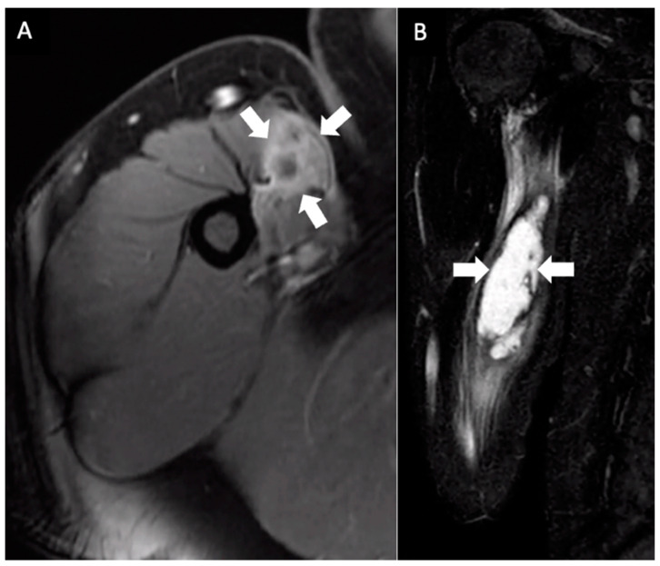
Figure 2.
Multiplanar MRI of the right upper arm taken one month after identifying the mass and seven months after receiving the booster dose. Axial fast spin gradient echo postcontrast (A) and coronal STIR (B) images show an ill-defined central fluid signal measuring 10 × 3.1 × 1.9 cm with prominent peripheral enhancement (arrows).

