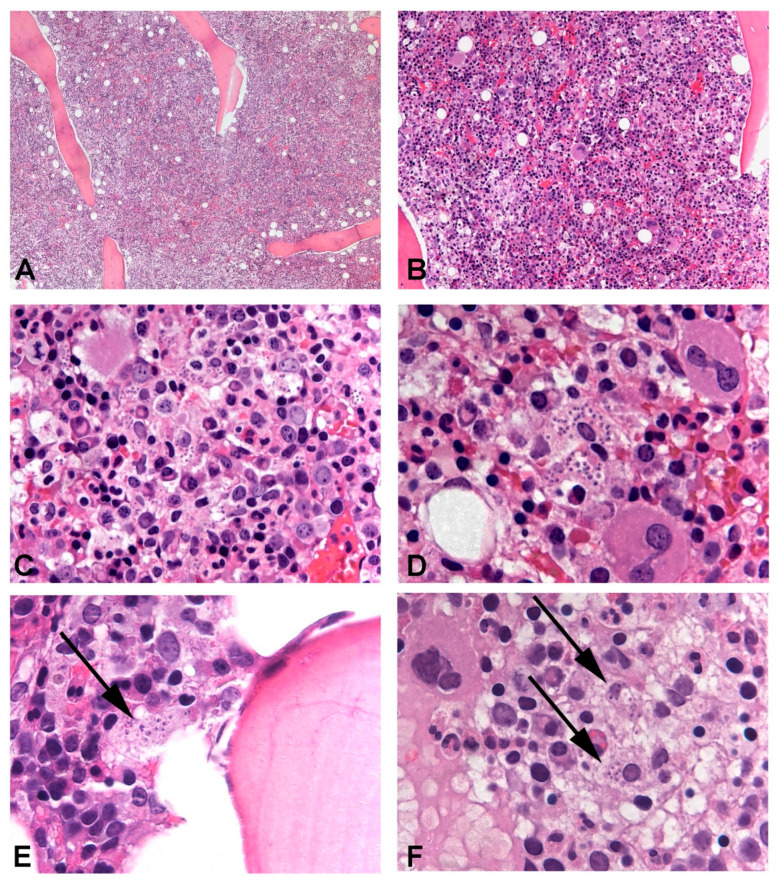
Figure 2.
Leishmania amastigotes could be identified in the bone marrow trephine of all patients, in variable amounts. In Case 3, intertrabecular lacunae were hypercellular and numerous macrophages phagocyting significant numbers of amastigotes were evident and could be easily observed (A–D). In Cases 1 and 2, parasitic infection was more subtle with only a few amastigotes identifiable in rare macrophages ((E) and (F), respectively). In Case 1, cellularity was increased, while in Case 2, it was normal and hematopoietic progenitors of the three series displayed signs of abnormal maturation.

