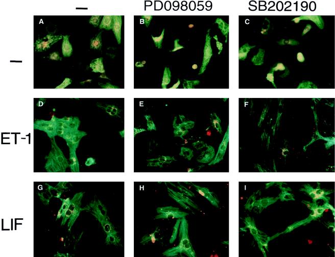
FIG. 5.
Inhibition of p38 activity changes the hypertrophic morphology induced by ET-1 and LIF in myocytes. Myocytes were either left untreated (A to C) or were treated with ET-1 (10 nM) (D to F) or LIF (1 nM) (G to I), in the presence or absence (−) of the specific MEK1 inhibitor PD098059 (50 μM) (B, E, and H) or the specific p38 inhibitor SB202190 (10 μM) (C, F, and I), as indicated. After 48 h, the cells were stained with anti-α-MHC monoclonal antibody, followed by fluorescein isothiocyanate-conjugated anti-mouse immunoglobulin (green). ANF polyclonal antibody was recognized by rhodamine-conjugated anti-rabbit immunoglobulin G (orange).

