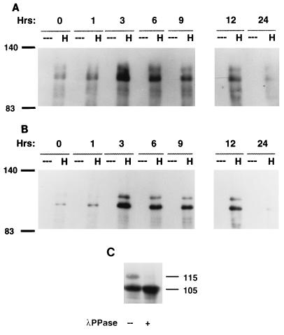
FIG. 5.
(A and B) p105HEF1 and p115HEF1 are tyrosine phosphorylated to comparable extents. Crude lysates were made from MCF-7 cells synchronized by thymidine block and released for the number of hours noted (0, 1, 3, 6, 9, 12, or 24). Lysates were immunoprecipitated either by control (---) or α-HEF1 (H) antibody. These lysates were resolved by SDS-PAGE and probed in Western blot analysis with the RC20 antibody to phosphotyrosine (A); following stripping, the blot was reprobed with α-HEF1 antibody (B). (C) p115HEF1 levels are reduced by treatment with lambda phosphatase. Lysates from HeLa cells transfected with pCMV-HEF1 were treated either with (+) or without (--) lambda phosphatase, resolved by SDS-PAGE, and visualized with α-HEF1. Numbers in the margins of each panel represent molecular mass in kilodaltons.

