FIG. 6.
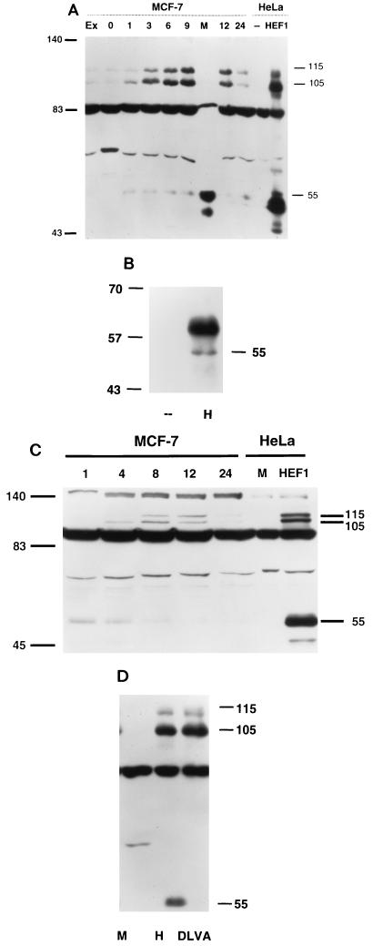
(A) Specific appearance of p55HEF1 in mitotic shakeoff. Crude lysates were made from either exponentially growing cells (Ex) or cells synchronized by thymidine block and released for the number of hours notes (0, 1, 3, 6, 9, 12, or 24). At 9 h following release, a mitotic shakeoff was prepared (M). As a control to indicate the position of the p105HEF1, p115HEF1 and p55HEF1 species, HeLa cell lysates (known to express relatively low levels of endogenous HEF1) either mock transfected (--) or transfected with pcDNA3-HEF1 (HEF1) were also analyzed. The lysates were resolved by SDS-PAGE and probed in Western blot analysis with α-HEF1 antibodies. (B) Endogenous p55HEF1 can be immunoprecipitated by antibody to HEF1. Five hundred micrograms of whole-cell lysate prepared from mitotic shakeoffs of MCF-7 cells 9 h after release from thymidine block was used for immunoprecipitation with either control (--) or α-HEF1 antibodies (H), followed by visualization with α-HEF1. Note that the prominent diffuse band migrating at ∼59 to 64 kDa represents the immunoglobulin blob generally detected in immunoprecipitations. (C) p55HEF1 is abundant in nocodazole-blocked cells and is replaced by p105HEF1 and p115HEF1 following release. MCF-7 cells were blocked in mitosis by incubation in 1 μM nocodazole for 14 h and released. Cell lysates were prepared from cells at 1, 4, 8, 12, and 24 h after release. As before, as a control for sizes, an aliquot of mock-transfected (M) or HEF1-transfected (HEF1) HeLa cells was included. Lysates were resolved by SDS-PAGE and probed in Western blot analysis with α-HEF1. (D) Production of p55HEF1 results from a cleavage of the full-length HEF1 protein at a DLVD motif located at aa 360 to 363. PCR-based mutagenesis was used to alter DLVD360–363 to DLVA in the context of the full-length 834-aa HEF1 coding sequence, and the mutant was cloned into the pCMV expression vector. Whole-cell lysates from HeLa cells mock transfected (M), transfected with pcDNA3-HEF1 (H), or transfected with pcDNA3-HEF1DLVA (DLVA) were visualized with α-HEF1 antibodies. Numbers in the margins of each panel indicate molecular mass in kilodaltons.

