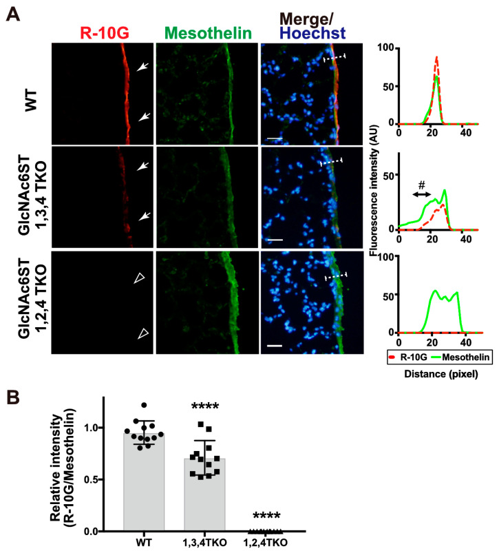
Figure 4.
GlcNAc6ST1,2,4 triple-KO mice lack R-10G-reactive sulfated glycans in the pleural mesothelium. (A) Lung sections prepared from normal wild-type (WT), Chst2/Chst5/Chst7 triple-deficient (GlcNAc6ST1,3,4 TKO), and Chst2/Chst4/Chst7 triple-deficient (GlcNAc6ST1,2,4 TKO) [28] mice were co-stained with R-10G (red) and an anti-mesothelin (green), followed by Hoechst 33342 nuclear staining (blue). Dense R-10G staining in the pleural mesothelium is demonstrated (arrows). GlcNAc6ST1,2,4 TKO mice showed negligible levels of R-10G signals in the mesothelium (open arrowheads). Plot profiles of R-10G and mesothelin staining are presented. Signal intensities along the line marker (white dashed line) paths in the merged images were determined (n = 3 per genotype). The area proximal to the basement membrane structure in the mesothelial cell layer showed a selective reduction in R-10G immunoreactivity (#). (B) The relative intensity of R-10G to mesothelin is indicated (n = 12 mesothelia per genotype). Data were obtained from three experiments in which four pleural mesothelia from lung specimens from three donors were analyzed for each genotype. **** p < 0.0001. Scale bar: 20 µm.

