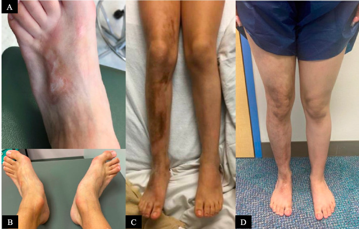
Fig. 4.
Features of localized scleroderma. A Patient 2’s damage features of localized scleroderma include visible venous pattern resulting from epidermal and dermal atrophy. There is a plaque of bound down, sclerotic skin causing hyperpigmentation, sclerotic bands, atrophy, and contractures. B As a sequela of fascial and tendon fibrosis as well as joint ankylosis, she also has bilateral severe flatfoot deformity with hindfoot valgus. C Patient 3’s initial diagnosis image including active right leg linear scleroderma with hyperpigmented plaques with central thickening surrounded by outer erythema. D Patient 3’s current physical examination shows late-stage morphea damage features including right lower extremity dermal atrophy with visible vessels, subcutaneous atrophy, dyspigmentation, decreased leg circumference, mild right ankle flexion contracture, and non-significant leg length difference (8 mm)

