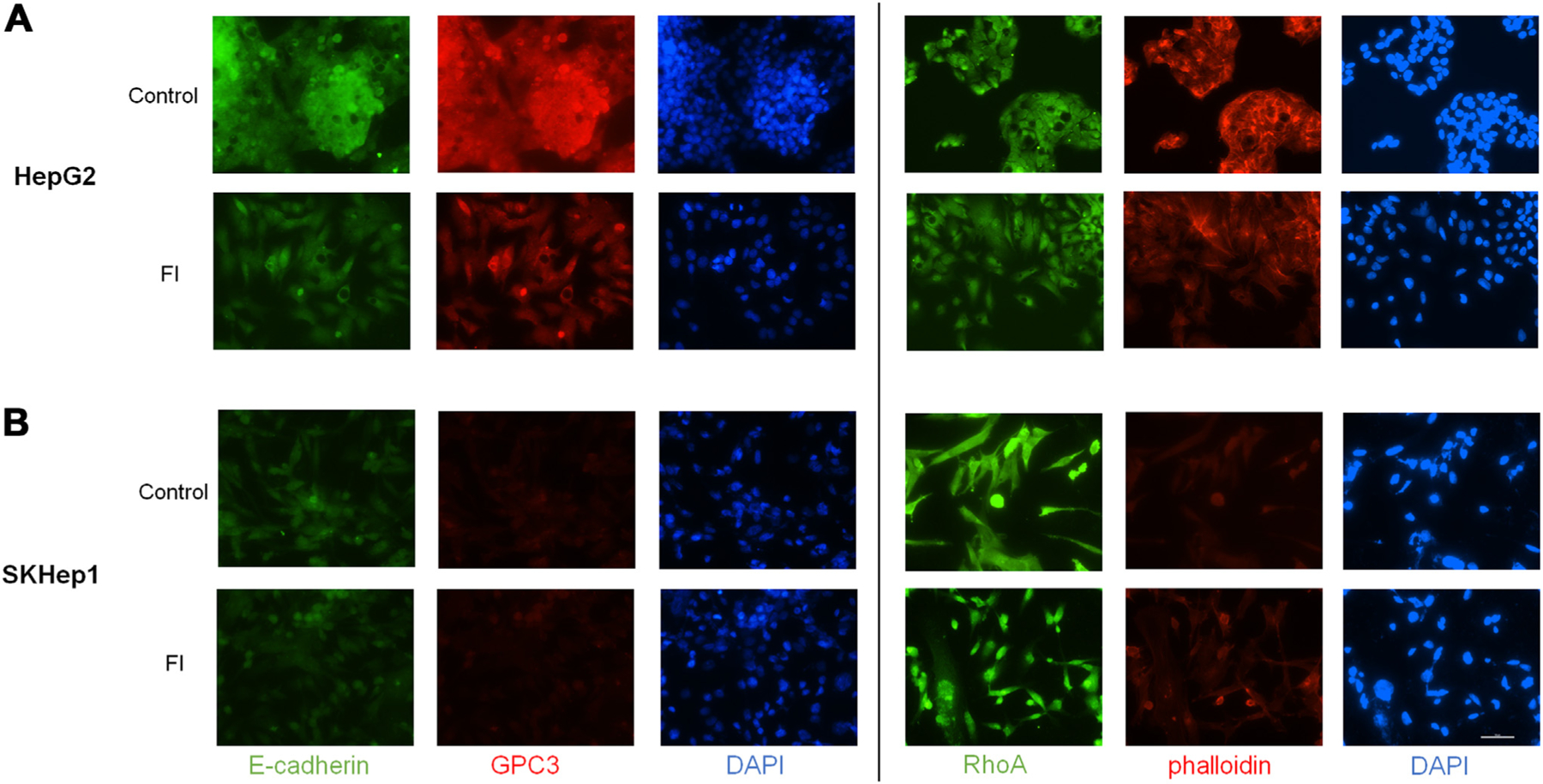
Fig. 3 –

Furin inhibition induces morphological changes and reduces expression of cytoskeletal and adhesion proteins. (A) HepG2 cells show reduced protein expression of E-cadherin as well as reorganization and potential reduction of total GPC3 at 24 h after FI treatment compared to control cells. RhoA and phalloidin after 24-h treatment with FI show reduction and reorganization. (B) No clear change in staining is observed in SKHep1 cells in FI treated cells compared to control although there may be some reorganization of RhoA. GPC3 staining is negative in SKHep1 cells as expected (20X objective, scale bar 100 μm).
