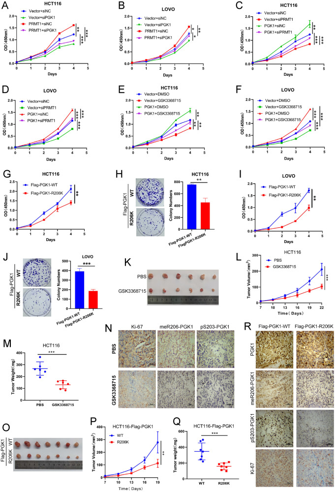
Fig. 5. PRMT1-mediated meR206-PGK1 promotes colorectal cancer cell proliferation and tumorigenesis.
A, B CCK-8 assays were used to assess the effect of PGK1 deficiency on cell proliferation in vector and PRMT1 stable overexpressed HCT116 cells (A) or LOVO cells (B). C, D CCK-8 assays were used to assess the effect of PRMT1 deficiency on cell proliferation in Vector and PGK1 stable overexpressed HCT116 cells (C) or LOVO cells (D). E, F CCK-8 assays were used to assess the effect of GSK3368715 on cell proliferation in vector and PGK1 stable overexpressed HCT116 cells (E) or LOVO cells (F). G, J CCK-8 assays and colony formation assays were used to assess the HCT116 cells (G, H) and the LOVO cells (I, J) proliferation abilities after stable over-expressing Flag-PGK1-WT or Flag-PGK1-R206K, respectively. K–N 4×10⁶ HCT116 cells and Matrigel (Corning; 1:1 ratio) were subcutaneously injected into the abdominal flanks of mice (n = 12). When the volume of the tumor reached about 50mm3, the mice were divided into two groups randomly: PBS and GSK3368715 (n = 6 for each group). The mice in the GSK3368715 group were treated with GSK3368715 100 mg/kg intraperitoneal everyday. Representative images (K), tumor volumes (L) and tumor weights (M) of the two groups were shown. IHC detected Ki-67, meR206-PGK1, and pS203-PGK1 expression in xenograft tumors (N). O–R 4×10⁶ stable overexpressed wild and mutant Flag-PGK1 (WT, R206K) HCT116 cells and Matrigel (Corning; 1:1 ratio) were subcutaneously injected into the abdominal flanks of mice (n = 7) to analyze the effect of meR206-PGK1 in tumorigenesis, representative images (O), tumor volumes (P) and tumor weights (Q) of the two groups were shown. IHC detected PGK1, Ki-67, meR206-PGK1, and pS203-PGK1 expression in xenograft tumors (R).

