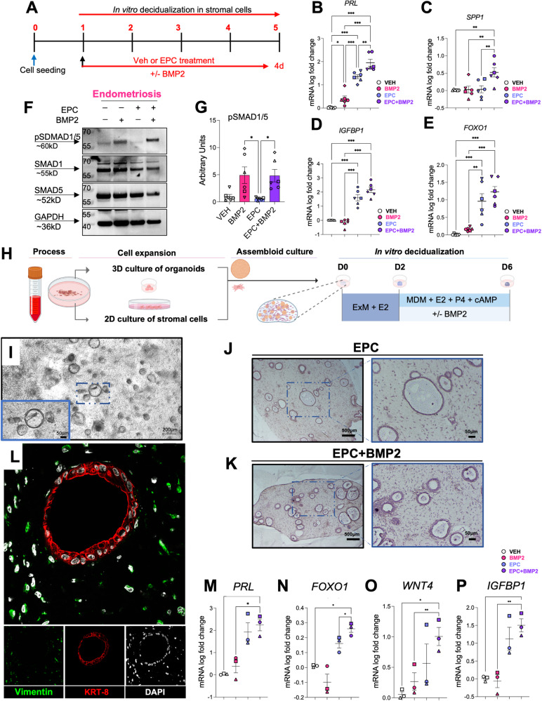
Fig. 6. BMP2 supplementation improves the decidualization potential of 2D and 3D endometriosis patient-derived endometrial cultures.
A Experimental outline showing the treatment groups used to test how the addition of recombinant BMP2 affects decidualization in EPC-treated stromal cells from individuals with endometriosis. B–E qRT-PCR quantification of decidualization markers PRL (B), SPP1 (C), IGFBP1 (D), and FOXO1 (E) following Vehicle, BMP2, EPC, or EPC + BMP2 treatment in stromal cells from individuals with endometriosis (n = 6). The different symbols represent individual patient trajectories per sample, plotted as mean +/− standard error of the mean. One-way ANOVA with a Tukey’s postdoc test. F, G Western blot analysis (F) and quantification (G) of endometrial stromal cells from individuals with endometriosis following 4 days of treatment with Vehicle, BMP2, EPC, or EPC + BMP2. H Diagram showing the experimental procedure for establishing endometrial epithelial and stromal co-cultures or “assembloids” from endometrial tissues of individuals with endometriosis. After the assembloids were established, they were pre-treated with 10 nM estradiol (E2) followed by decidualization with the EPC decidualization cocktail (1 µM MPA, 0.5 mM cAMP and 1 µM E2) +/− 25 ng/ml BMP2 for an additional 4 days. Image was created using BioRender. I Phase contrast micrograph of the endometrial epithelial and stromal assembloids showing the endometrial epithelial organoids and the distribution of stromal cells in the collagen matrix. J–L Histological analysis of cross sections obtained from the endometrial assembloids stained with hematoxylin and eosin (J, K) or using immunofluorescence using vimentin (green), cytokeratin 8 (KRT-8, red) or DAPI (white) (L). M–P qRT-PCR analysis of decidualization markers, PRL (M), FOXO1 (N), WNT4 (O), or IGFBP1 (P) in the endometrial assembloids treated with Vehicle, BMP2, EPC, or EPC + BMP2. Plotted values represent mean +/− standard error of the mean, with the different symbols corresponding to each patient’s trajectory. Data were analyzed using a one-way ANOVA with a Tukey’s posthoc test.

