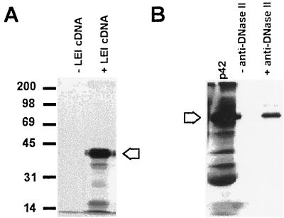
FIG. 3.
Expression of porcine LEI in vitro. (A) The cDNA of porcine LEI was inserted in the pGEM vector and expressed with Promega reticulocyte lysate. The reaction was allowed to proceed for 1.5 h at 30°C in the presence or absence of plasmid DNA. A 3-μl volume of reaction mixture was mixed with the same volume of 2× Laemmli sample buffer. The samples were separated on a 12% acrylamide gel and treated for autoradiography. The arrow indicates the 42-kDa main band. (B) A 5-μl volume of reticulocyte lysate containing [35S]methionine-labelled LEI was immunoprecipitated in the presence or absence of anti-DNase II and then analyzed by PAGE and autoradiography. The arrow indicates the 42-kDa band.

