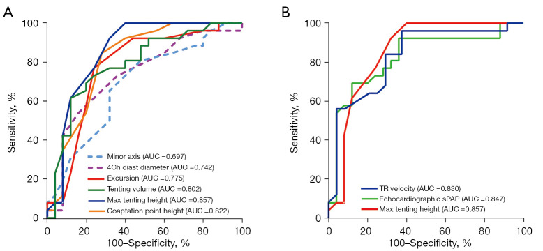
Figure 4.
ROC curves of conventional echocardiography and 4D echocardiography to detect severe PH in high-quality image cases. (A) ROC curves of minor axis, 4Ch diastolic diameter, excursion, tenting volume, MTH, and coaptation point height to detect severe PH. (B) ROC curves of TR velocity, echocardiography sPAP, and MTH to detect severe PH. AUC, area under the curve (P<0.05 for all). AUC, area under the curve; TR, tricuspid regurgitation; sPAP, systolic pulmonary arterial pressure; ROC, receiver operator characteristic; 4D, 4-dimensional; MTH, maximal tenting height; PH, pulmonary hypertension.

