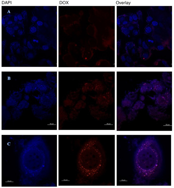
Figure 8.
Confocal images of resistant MCF-7. (A) Confocal imaging of control resistant MCF-7/DOX 26.6 nM cell line. (B) Confocal imaging of treated resistant MCF-7 cell line, the free DOX surrounding the nucleus and also presented at (C) a higher magnification of image B. Magnification, 20X. DOX, doxorubicin.

