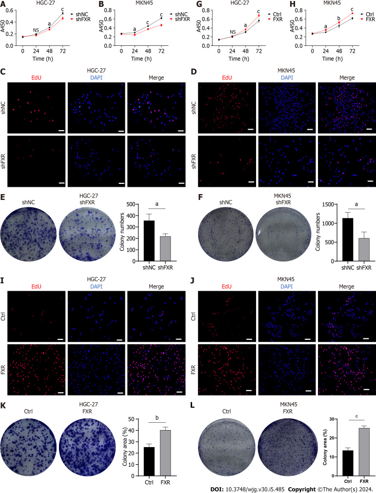
Figure 6.
Farnesoid X receptor promoted proliferation of gastric cancer cells. A-F: Malignant proliferation assays, including cell viability (A and B), 5-ethynyl-2′-deoxyuridine (Edu) staining (C and D), andcolony formation assays (E and F), were performed in gastric cancer (GC) cells after transfection with the shNC or shFXR plasmid; G-L: Cell viability (G and H); Edu staining (I and J), andcolony formation assays (K and L) were performed in GC cells after transfection with the control or farnesoid X receptor-coding plasmid. Scale bar: 100 μm. aP < 0.05, bP < 0.01, cP < 0.001. These experiments were repeated three times. FXR: Farnesoid X receptor; Edu: 5-ethynyl-2′-deoxyuridine; NC: Negative control; NS: Not significant.

