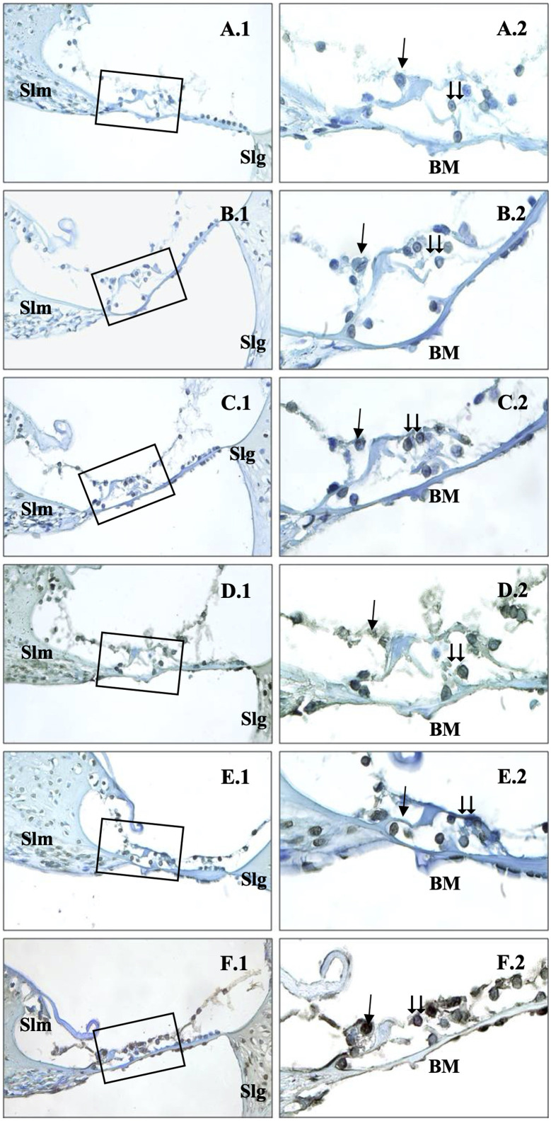
Figure 5.
Immunohistochemical staining of cochlea with TUNEL. →: inner hair cells, ⇉: outer hair cells, Slm: spiral limbus, Slg: spiral ligament, BM: Basillary membrane, Control (A), 0.6 V/m EF (B), 1.9 V/m EF (C), 5 V/m EF (D), 10 V/m EF (E), 15 V/m EF (F). Organ of Corti ×40 (1) and organ of Corti ×100, methyl green base staining (2).

 Content of this journal is licensed under a