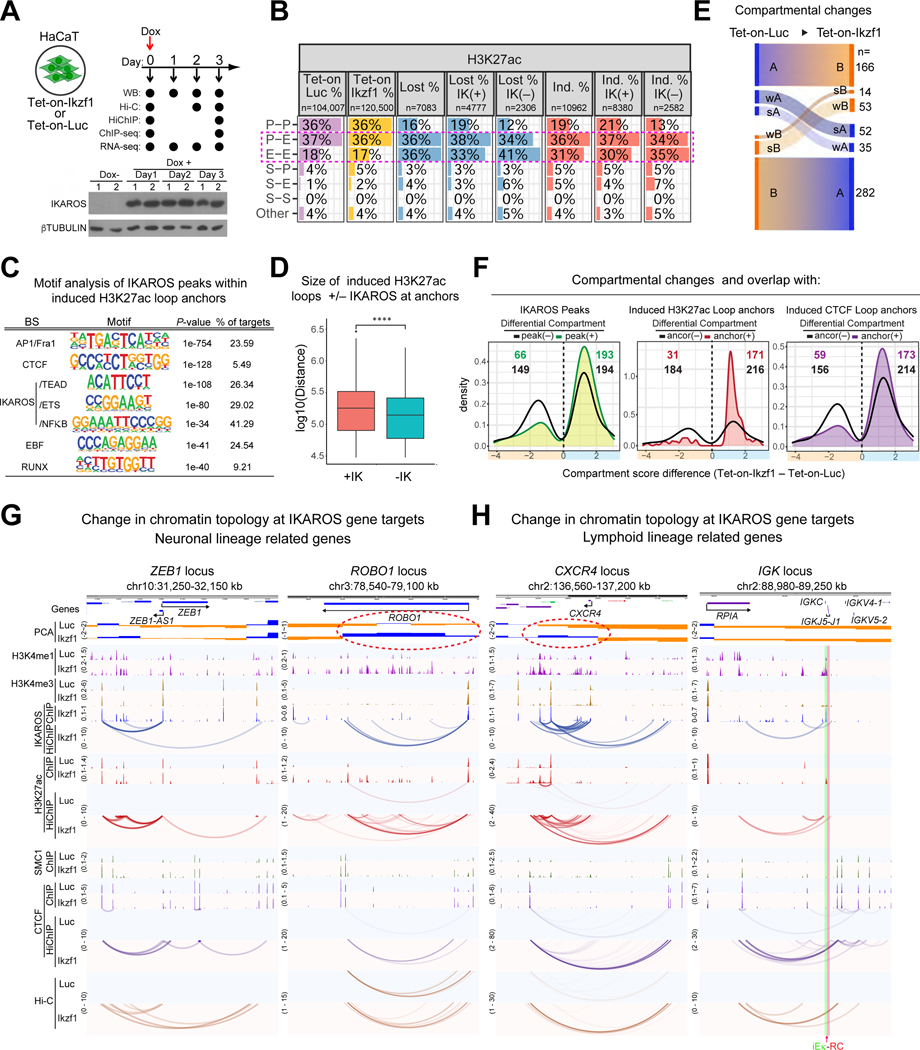
Figure 7. Extra-lineage chromatin organization induced by IKAROS in skin epithelial cells.
(A) Experimental strategy and Western blot of IKAROS induction. (B) Number and percentage of loops and differential loops (Lost, Induced (Ind.)) from H3K27ac HiChIP at d3 stratified according to anchor classification and presence/absence of IKAROS ChIP-seq peaks in at least one anchor (IK(+), IK(−)). (C) De novo TF-binding motif enrichment of IKAROS peaks within upregulated H3K27ac loop anchors. (D) Size distribution of induced H3K27ac loops at d3 with (+IK) or without (-IK) IKAROS at their anchors. **** p <= 0.0001. (E) Compartmental changes between Luciferase- and IKAROS-expressing cells at d2. (F) Distributions of differential compartmental regions with and without overlap with IKAROS peaks or induced H3K27ac or CTCF loop anchors. (G-H) Examples of IKAROS-mediated induction of chromatin topology. The IGK RC (red) and iEκ (green) are highlighted. Source: Tables S3-4, S6-7.

