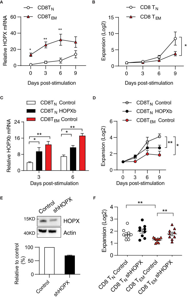
Figure 3.
HOPX contributes to the proliferation difference from human CD8+TN to TEM cells in response to stimulation. (A) Higher HOPX mRNA level in CD8+ TEM than in CD8+ TN cells before and after in vitro stimulation. CD8+ TN (CD62L+CD45RA+) and TEM (CD62L-CD45RA-) cells were isolated from healthy donors by cell sort and stimulated (anti-CD3/CD28) and harvested at day 0, 3, 6, and 9. The expression of HOPX mRNA in different T cell subsets was measured by qRT-PCR and normalized to the level of ACOX1. The mean ± SEM are present (n=5). Two-way ANOVA was used to calculate p value between two conditions. (B) Growth of CD28+ CD8+ TN and CD28+ CD8+ TEM cells in vitro post stimulation (anti-CD3/CD28). Stimulated cells were counted at the indicated days and the mean ± SEM are present (n=7). (C) HOPX mRNA level in HOPXb and control transduced CD8+ TN, and control transduced TEM cells in vitro. TdTomato+ cells were isolated by cell sorter from HOPX over-expressing or control lentiviral transduced CD8+ TN, and TEM cells. HOPX mRNA level was measured in control CD8+TN, CD8+TEM, and HOPXb over-expressing CD8+TN cells by qRT-PCR at day 3 and 6 post stimulation and normalized to ACOX1. The mean ± SEM are present (n=3-4). (D) Growth of HOPXb over-expressing CD8+TN cells and control CD8+TN and TEM cells in vitro. Cell numbers were counted based on tdTomato positive cells at day 3, 6, and 9. The mean ± SEM are present (n=5). Two-way ANOVA was used to calculate p value between two conditions. (E) Reduced HOPX protein by shRNA knockdown. A shRNA (target on exon2 of HOPX) was designed and cloned into a lentiviral vector and then transduced Jurkat cell line and the efficiency of shRNA-mediated knockdown was confirmed by Western blot. A representative graph (Western blot) showing the level of HOPX in HOPX-transduced Jurkat cells 3 days after treatment with either control or HOPX shRNA and quantification (20-30% reduction). (F) Increased growth of CD8+TEM cells post HOPX knockdown. CD8+TEM and CD8+TN cells were isolated, stimulated, and transduced with HOPX shRNA and control lentiviruses. Cell numbers were counted at day 2 post viral transduction and the mean ± SEM are present (n=9) with p value using Student’s t-test. * as p≤ 0.05, ** as p≤ 0.01.

