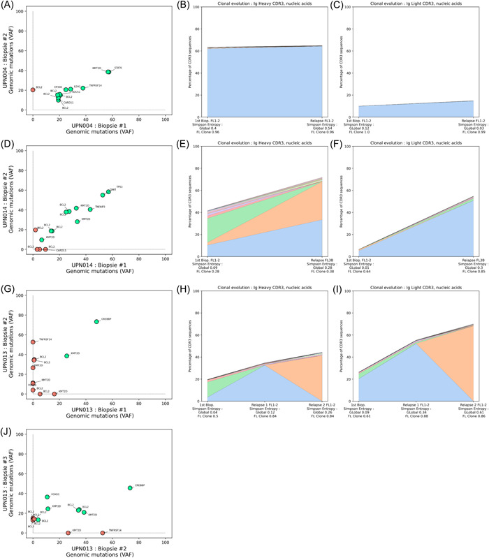
Figure 7.

Evolution of contributions of the major follicular lymphoma (FL) subclones and genetic mutation patterns during FL progression. Three representative cases are presented, and all cases with paired samples are available in Figure S4. The variant allele frequencies of the detected somatic mutations identified in genomic DNA were plotted at diagnosis and relapse to assess the evolution of the FL population (A, D, G, and J), with a green circle for concordant mutation between diagnosis and relapse, and a red circle for discordant mutations. The evolutions of the dominant FL subclones were evaluated in parallel through the analysis of the Ig CDR3 sequences, representing the contribution of the 10 most frequent sequences for the IgH (B, E, and H) and IgL (C, F, and I) loci.
