
FIG. 2.
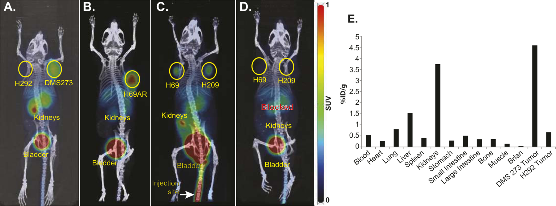
PET/CT imaging using 68Ga-pentixafor demonstrates specific targeting of CXCR4 in SCLC. The mice showed the expected biodistribution with radiotracer uptake seen in CXCR4-expressing tumors as well as excretion via the kidneys and bladder. In Panels A–D, images of mice bearing SCLC xenografts were taken 1 h after injection with 168–334 μCi 68Ga-pentixafor. PET images were set with a scale of 0–1. Panel A: H292 NSCLC xenograft was used as a negative control and DMS273 SCLC xenograft; Panel B: H69AR SCLC xenograft; Panel C: H69 and H209 SCLC; Panel D: Same mouse as Panel C with 50 μg AMD3100 (unlabeled CXCR4 antagonist) co-injected with 68Ga-pentixafor; Panel E: Bio-distribution of 68Ga-pentixafor in mouse shown in Panel A, measured ~1 h 45 min after injection.