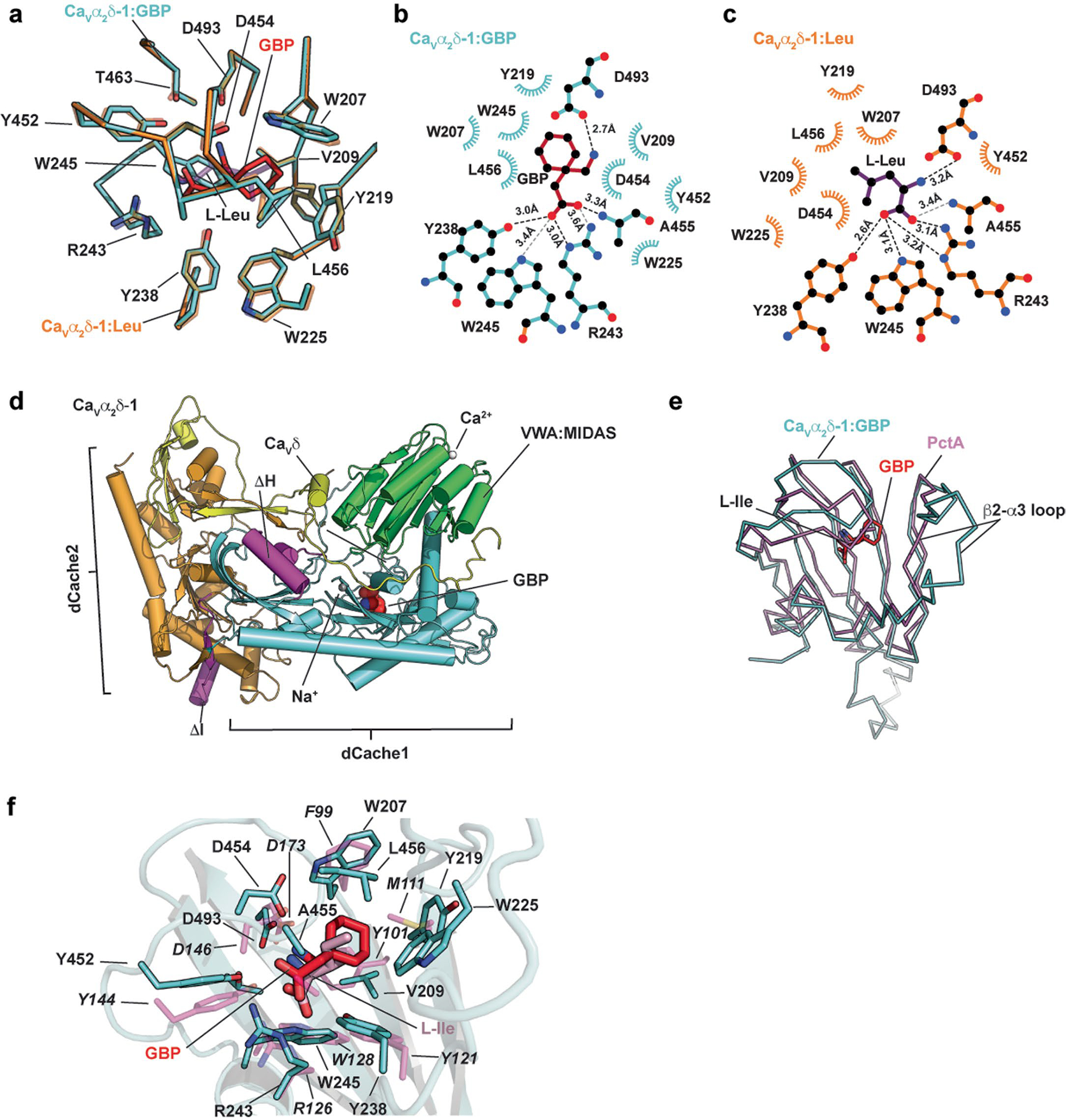
Extended Data Fig. 4 |. CaVα2δ-1 GBP-binding site analysis and comparisons.

a, Superposition of the CaVα2δ-1:GBP (aquamarine) and CaVα2δ-1:l-Leu (orange) (PDB:8EOG)13 binding sites. GBP is red. l-Leu is purple. b and c, LigPLOT37 diagrams of the b, CaVα2δ-1:GBP (aquamarine) and c, CaVα2δ-1:l-Leu (orange) (PDB:8EOG)13 binding sites showing hydrogen bonds and ionic interactions (dashed lines) and van der Waals contacts ≤ 5 Å. GBP is red. l-Leu is purple. d, Superposition of the first dCache1 repeats from CaVα2δ-1:GBP (aquamarine) and the PctA:l-Ile complex (magenta) (PDB: 5T65)34. GBP is red. e,f, Closeup view of superposition from ‘d’ showing ligand contact residues. CaVα2δ-1 is shown as a cartoon. GBP is red. Corresponding sidechains of PctA are magenta. l-Ile form the PctA complex is pink. PctA residues are labeled in italics.
