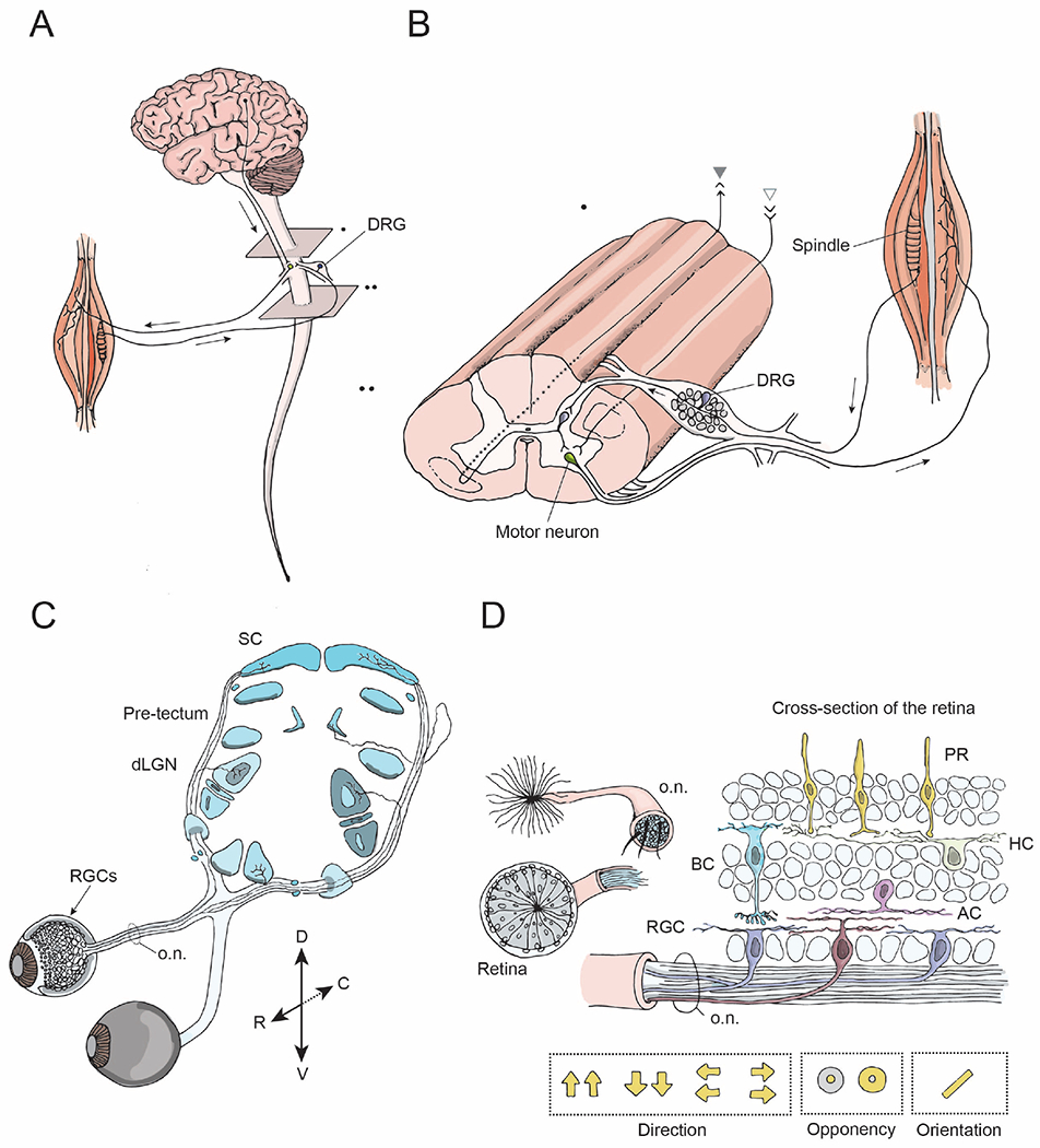
Figure 1: Model systems for studying CNS regeneration: spinal system and visual system.

(A) The brain and the spinal cord exchange information via descending projections from the brain to the spinal cord (arrow), and ascending projections. The spinal cord receives sensory input from external stimuli via the dorsal root ganglia (DRG) neurons. Output signals from the brain and the spinal cord are relayed to the muscles via motor neurons and their peripheral projections.
(B) A longitudinal view of the spinal cord showing sensory inputs, interneurons, and motor outputs. The spinothalamic tract is comprised of ascending projections from interneurons (gray, closed arrowhead), whereas the corticospinal tract consists of descending projections from the brain to the spinal cord (gray, open arrowhead). The DRG neurons have central and peripheral axonal branches.
(C) The visual system includes retinal ganglion cells (RGCs, arrow), the neurons that relay sensory input from the eye, their axons that form the optic nerve (o.n.) and their projections to target regions within the brain including the dorsal lateral geniculate nucleus (dLGN) and the superior colliculus (SC) (blue).
(D) Cross-section of the retina and the optic nerve. The retina consists of five types of neural cells: photoreceptors, i.e. rods and cones (PR, yellow), bipolar cells (BC, blue), horizontal cells (HC, green), amacrine cells (AC, pink) and ~40 different types of retinal ganglion cells (RGC, brown, purple). RGCs are the only neurons that send projections from the retina into the optic nerve and have various receptive field properties as indicated by the yellow arrows and circles.
