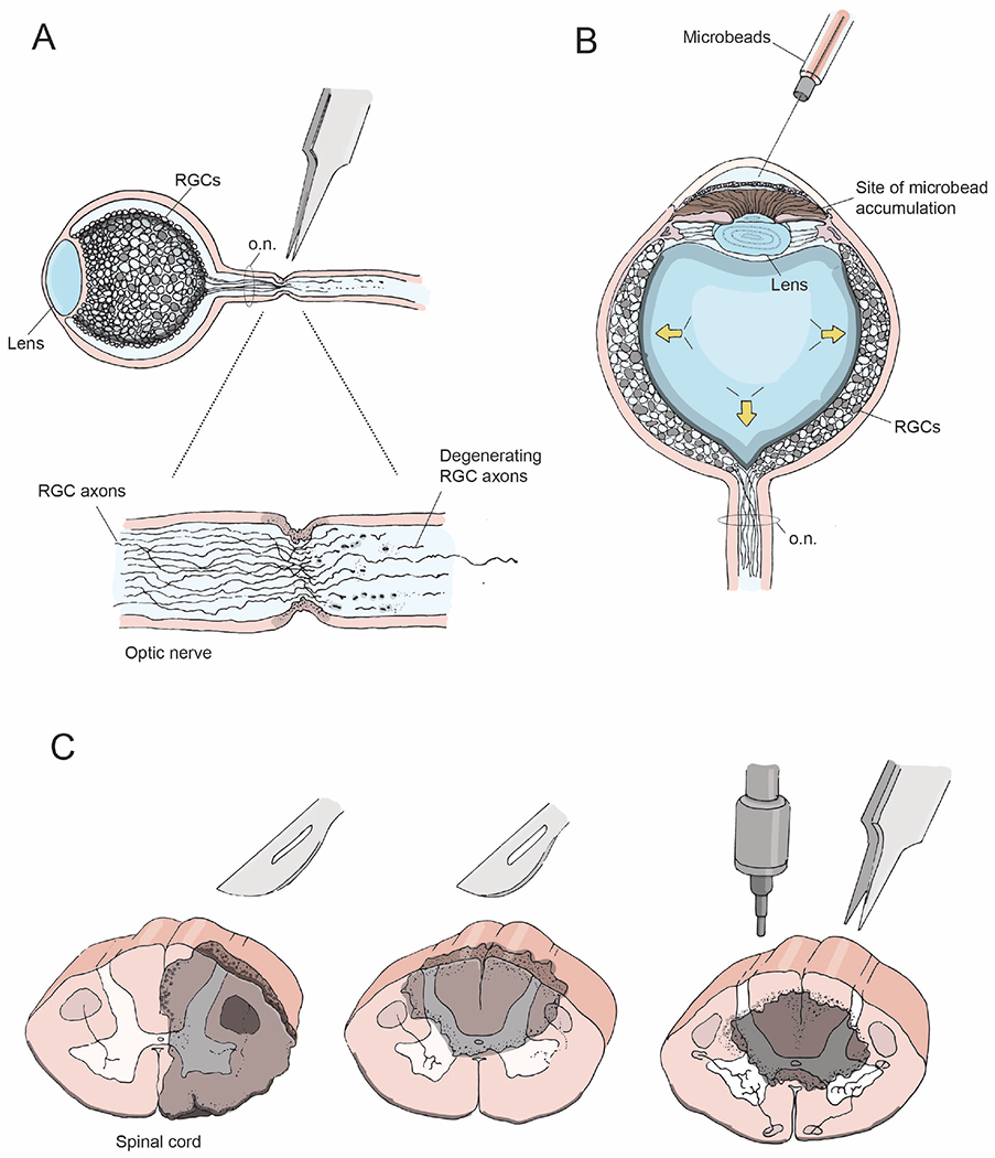
Figure 2: CNS injury models.

(A) Optic nerve crush injury showing axon degeneration distal to the injury site
(B) Bead-induced mouse model of glaucoma, wherein microbeads injected into the eye increase intraocular pressure (arrows) and mimic the degenerative effects of glaucoma
(C) Coronal views of the spinal cord depicting lesion sites (gray) following unilateral transection (left), dorsal bilateral hemisection (middle), and a contusion or crush injury (right).
