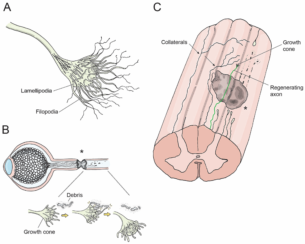
Figure 3: Axon growth following injury.

(A) Growth cone, i.e. the leading edge of an axon. The fingerlike protrusions are filiopodia and lamellipodia that are composed of actin filaments and are crucial for the growth cone’s ability to grow towards or away from environmental cues.
(B) Axons regenerating following an optic nerve crush injury. Growth cones at the leading edge of regenerating axons grow past the lesion site (asterisk), interact with microglia/axonal-debris/guidance cues, and accordingly alter their direction of growth.
(C) Longitudinal view of the spinal cord showing a lesion site (gray), distal processes of injured axons degenerating (dotted lines), a regenerating axon extending a growth cone distal to the lesion site (color), and two spared axons extending collaterals circumventing the lesion site and extending to target neurons distal to the lesion (arrows).
