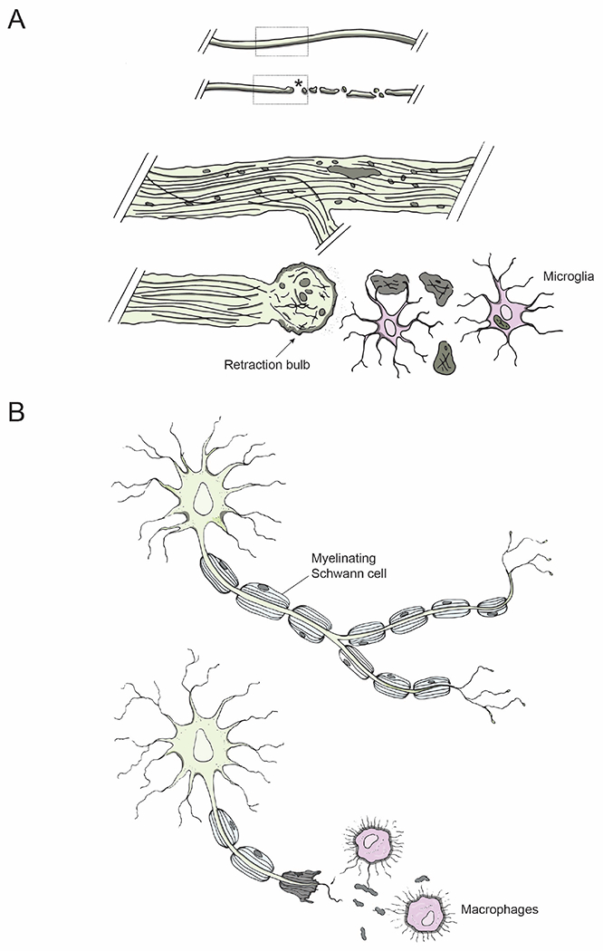
Figure 4: Axon degeneration and immune response to injury.

(A) Degenerative mechanisms following injury: An intact axon before injury; an injured axon undergoing Wallerian degeneration distal to the lesion site (asterisk). Dotted regions of the intact and injured axons are shown magnified below: retraction bulb (arrow) sealing the axolemma at the proximal end of an injured axon and microglia (pink) clearing axonal debris.
(B) Injury in the PNS: Schwann cells in the PNS myelinate regenerating axons (top); macrophages phagocytose axonal debris (bottom, pink).
