
Figure 6.
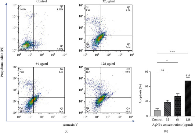
(a) Analysis of flow cytometry related to SKBR3 cells treated with silver nanoparticles for 24 h, obtained from Annexin-V/PI staining. Q1 quadrant shows necrotic cells stained by PI only, Q2 demonstrates late apoptotic cells, Q3 quadrant is early apoptotic, and Q4 quadrant contains viable cells. (b) Apoptosis rate of SKBR3 cells treated with silver nanoparticles which indicate increased apoptosis in 128 μg/ml. The results are shown as mean ± SEM in three replicates (n = 3). ∗P < 0.05; ∗∗,##P < 0.01; ∗∗∗P < 0.001. ns: nonsignificant changes. ∗Compared to the control group. #Compared to the 32 μg/ml group.