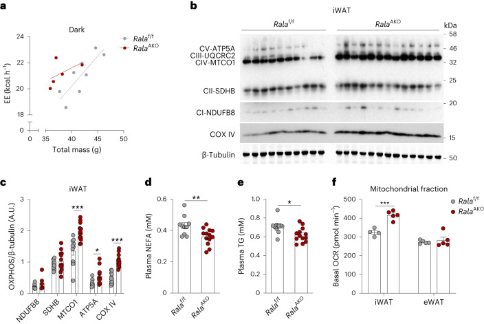
Fig. 3. RalA deficiency in WAT increases energy expenditure and mitochondrial oxidative phosphorylation.
a, Regression plot of energy expenditure (EE) measured in HFD-fed Ralaf/f (n = 8) and RalaAKO (n = 5) mice during dark phase. ANCOVA was performed using body weight (BW) as a covariate, group effect P = 0.0391. b,c, Immunoblot (b) and quantification (c) of OXPHOS complex proteins and β-tubulin in iWAT of HFD-fed Ralaf/f (n = 10) and RalaAKO (n = 13) mice. P = 0.0005, P = 0.0348, P < 0.0001. d,e, Plasma non-esterified fatty acid (NEFA; d) and TG (e) levels in HFD-fed Ralaf/f (n = 10) and RalaAKO (n = 13) mice; P = 0.0077(d), P = 0.0115 (e). f, Basal OCR in mitochondria measured by Seahorse. Mitochondrial fractions were isolated from primary mature adipocytes in iWAT or eWAT of HFD-fed Ralaf/f (n = 4) and RalaAKO (n = 5) mice. iWAT P = 0.0004. Data (c–f) show mean ± s.e.m., *P < 0.05, **P < 0.01, ***P < 0.001 by two-tailed Student’s t-test (c–f).

