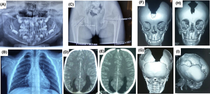
FIGURE 2.

Radiological features of cleidocranial dysplasia. (A) The orthopantomogram at age 8 showing germ retention and polyodontia with supernumerary teeth; (B) Chest X‐rays showing a total absence of the clavicles and (C) Pelvic x‐rays showing a left coxa Vara lesion; Brain CT‐scan showing (D) a dilatation of the occipital horns of the lateral ventricles at the time of diagnosis and (E) 3 years later; 3D reconstruction of the CT‐scan of the skull in (F) anterior view showing patent anterior fontanel, the absence of the upper part of the frontal and the parietal bones, (G) posterior view showing the absence of the parietal bone with preservation of the occipital bone, and (H, I) progression of the cranial bone development after 3 years of evolution with a notable reduction of the bone defect.
