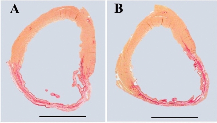
Fig. 2.
Representative slices of hearts stained with picrosirius red staining. Muscle cells stain yellow while collagen (scar) stains red. A Air-exposed heart with myocardial infarct scar as control; B E-cig-exposed heart with myocardial infarct scar. Note that scar circumference was comparable in the air-exposed heart compared to the E-cig-exposed heart, and the scar thickness and total LV circumference were also comparable between the two groups. (Scale bar = 5 mm)

