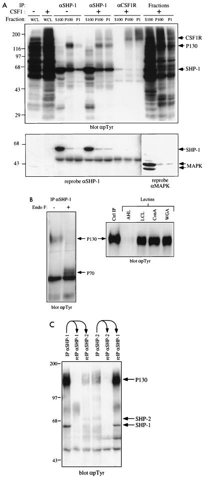
FIG. 1.
P130 is a membrane-localized glycoprotein and associates with both SHP-1 and SHP-2. (A) SHP-1–P130 complex localizes to the membrane compartment. BAC1.2F5 cells were starved of CSF-1 for 20 h and then stimulated with 2,000 U of CSF-1 per ml for 1 min (+) or left unstimulated (−). Cells were fractionated as described in Materials and Methods, and aliquots of the P1, P100, and S100 fractions were immunoprecipitated (IP) with anti-SHP-1 (αSHP-1) or anti-CSF-1R antibodies. Immune complexes were analyzed by SDS-PAGE (10% gel) and anti-pTyr immunoblotting. The anti-SHP-1 antibodies used for this panel are weakly cross-reactive with SHP-2, accounting for the 70-kDa phosphotyrosyl species; SHP-1-specific reagents are used in all subsequent experiments. Whole-cell lysates (WCL) and each fraction also were analyzed. Blots were reprobed with an anti-SHP-1 MAb to test for protein levels, and blots of each fraction were reprobed with anti-MAPK to monitor contamination of the P1 and P100 fractions with soluble cytoplasmic proteins. The positions of migration of Gibco BRL protein molecular size standards are shown in kilodaltons at the left. (B) P130 is a glycoprotein. Anti-SHP-1 immunoprecipitates from randomly growing BAC1.2F5 cells were treated with endo F as described in Materials and Methods. Deglycosylated (+) and untreated (−) samples were analyzed by SDS-PAGE (8% gel) and anti-pTyr immunoblotting (left panel). Lectin binding assays (right panel) were carried out as described in Materials and Methods, using A. hypogaea lectin (AHL), L. culinaris lectin (LCL), concanavalin A (ConA), or wheat germ agglutinin (WGA). Bound proteins were analyzed by SDS-PAGE (8% gel) and anti-pTyr immunoblotting. An anti-SHP-1 immunoprecipitate was loaded as a control (Ctrl IP). (C) P130 can coimmunoprecipitate with SHP-1 and SHP-2. Specific antibodies to SHP-1 and SHP-2 were used in consecutive immunoprecipitations from the lysates of a randomly growing normal macrophage cell line (N). Bound proteins were analyzed by SDS-PAGE (8% gel) and anti-pTyr immunoblotting. The migration of molecular size standards is indicated in kilodaltons at the left.

