Fig. 1.
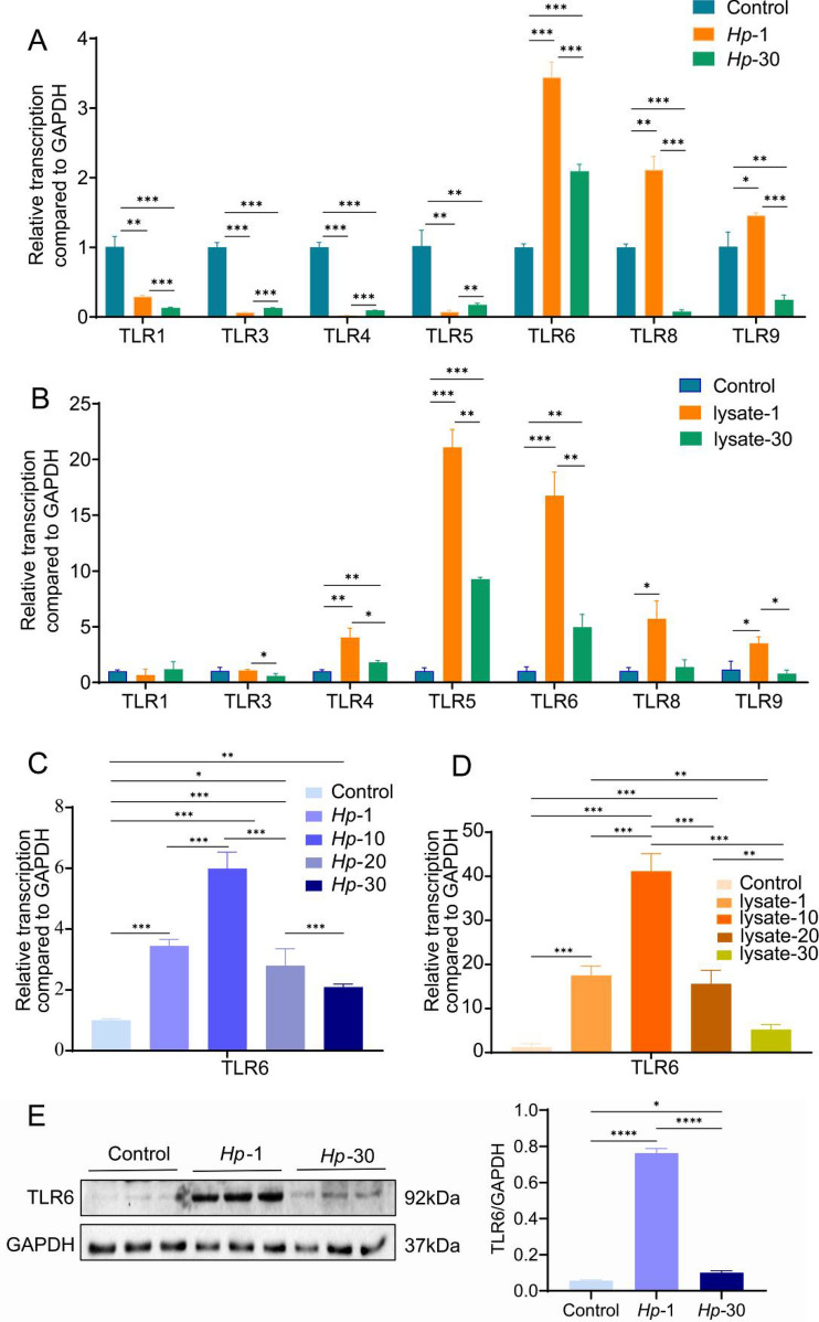
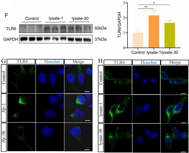
TLRs mRNA expression in (A) wild-type GES-1 cells (control), GES-1 cells persistently infected with H. pylori for 1 generation (Hp-1) and 30 generations (Hp-30) or in (B) control cells, GES-1 cells co-cultured with H. pylori lysate for 1 generation (lysate-1) and 30 generations (lysate-30). TLR6 mRNA expression in (C) control cells, Hp-1 cells, Hp-10 cells, Hp-20 cells and Hp-30 cells or in (D) control cells, lysate-1 cells, lysate-10 cells, lysate-20 cells and lysate-30 cells. TLR6 protein expression (E) and TLR6 immunofluorescent staining (G) in control cells, Hp-1 cells and Hp-30 cells. TLR6 protein expression (F) and TLR6 immunofluorescent staining (H) in control cells, lysate-1 cells and lysate-30 cells. All experiment were performed in triplicate and all data represents means ± SEM. ***P < 0.001, **P < 0.01, *P < 0.05. Scale bars, 10 µm (G, H)


