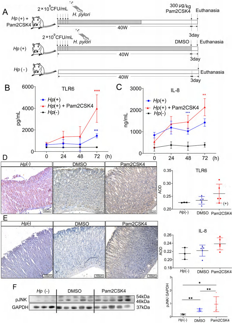
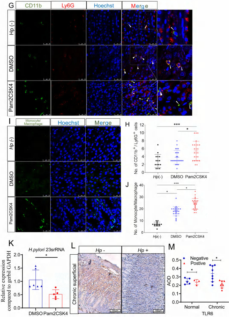
Fig. 6.
Schematic (A) of the in vivo treatment experimental plan. Serum TLR6 and IL-8 expression (B, C) and gastric mucosa TLR6 and IL-8 IHC staining (D, E) of the H. pylori-negative Mongolian gerbils or in the H. pylori-infected gerbils after 24 h, 48 h and 72 h intraperitoneal administration of Pam2CSK4 or DMSO. pJNK expression (F) in the gastric mucosa of H. pylori-negative Mongolian gerbils or in the H. pylori-infected gerbils after 72 h intraperitoneal administration of Pam2CSK4 or DMSO. The Immunofluorescence staining of CD11b+/Ly6G+ cells (G), monocyte/macrophage (I), the number of CD11b+/Ly6G+ cells (H) and the number of monocyte/macrophage (J) in gastric mucosa of H. pylori-negative gerbils, H. pylori-positive gerbils treated with DMSO or Pam2CSK4. The level of H. pylori 23srRNA (K) in gastric mucosa of H. pylori-positive Mongolian gerbils treated with DMSO or Pam2CSK4. IHC staining of TLR6 expression (L) in the gastric mucosa of patients. × 100. The average optical density (AOD) of TLR6 expression (M) in patients’ gastric mucosa. All experiment were performed in triplicate and all data represents means ± SEM. ***P < 0.001, **P < 0.01, *P < 0.05


