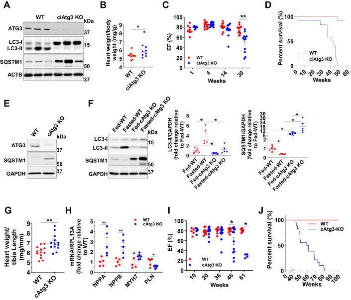
Figure 1. Cardiac-specific ATG3 deletion blocks autophagy and precipitates cardiac contractile dysfunction.
(A) ATG3, LC3 (MAP1LC3A), SQSTM1, and beta-actin (ACTB) protein levels in WT and ciAtg3 KO mouse hearts 1 week after last tamoxifen injection. Mice, at 6 weeks of age, were intraperitoneally injected with tamoxifen at a dose of 100 µg/g body weight/day for 5 consecutive days. One week after last tamoxifen injection, mice were euthanized and hearts were harvested for experiments. Age-matched ATG3loxP/loxP mice without Cre were injected with the same amount of tamoxifen as WT control. Representative blots are shown. n = 3 per group. (B) Heart weight of WT and ciAtg3 KO mice, 1 week after last tamoxifen injection. n = 7–8. Data are mean ± SEM. An unpaired t test was used to determine statistical significance between two groups. *P < 0.05. (C) Ejection fraction in WT and ciAtg3 KO mice 1, 4, 14, and 30 weeks after last tamoxifen injection. Measurements were performed under light sedation with midazolam. n = 7–12 per group. Data are mean ± SEM. Unpaired t tests were used to determine statistical significance between two groups at corresponding time points. **P < 0.01. (D) Survival curve of WT and ciAtg3 KO mice after the last tamoxifen injection. n = 12 per group. (E) ATG3, SQSTM1, and GAPDH protein levels in cardiomyocytes. Cardiomyocytes were enzymatically isolated from WT and cAtg3-KO mouse hearts. Representative blots are shown. This experiment was repeated twice independently. (F) LC3, SQSTM1, and GAPDH protein levels in mouse hearts. Mice were either randomly fed or fasted for 48 h, n = 5 per group. Data are mean ± SEM. One-way ANOVA followed by Bonferroni’s multiple comparison tests was used to determine statistical significance. *P < 0.05. (G) Heart weight of WT and cAtg3-KO mice at 16 weeks of age. n = 13 per group. Data are mean ± SEM. An unpaired t test was used to determine statistical significance between two groups. **P < 0.01. (H) NPPA, NPPB, MYH7, and PLN mRNA expression levels in WT and ATG3 KO mouse hearts at 16 weeks of age, n = 5 per group. Unpaired t tests were used to determine statistical significance between two groups. Data are mean ± SEM. **P < 0.01. (I) Ejection fraction in WT and cAtg3-KO mice at ages as indicated. Measurements were performed under light sedation with midazolam. n = 4–12 per group. Unpaired t tests were used to determine statistical significance between two groups at corresponding time points. Data are mean ± SEM. *P < 0.05. (J) Survival curve of WT and cAtg3-KO mice. n = 18. Data information: Cardiomyocytes for (E) were enzymatically isolated from 16-week-old WT and cAtg3-KO mice at a time when cardiac function was preserved in cAtg3-KO (I). After being enzymatically isolated, cardiomyocytes were immediately lysed in ice-cold homogenization buffer without any pre-treatments or culture. This experiment was repeated twice independently. Source data are available online for this figure.

