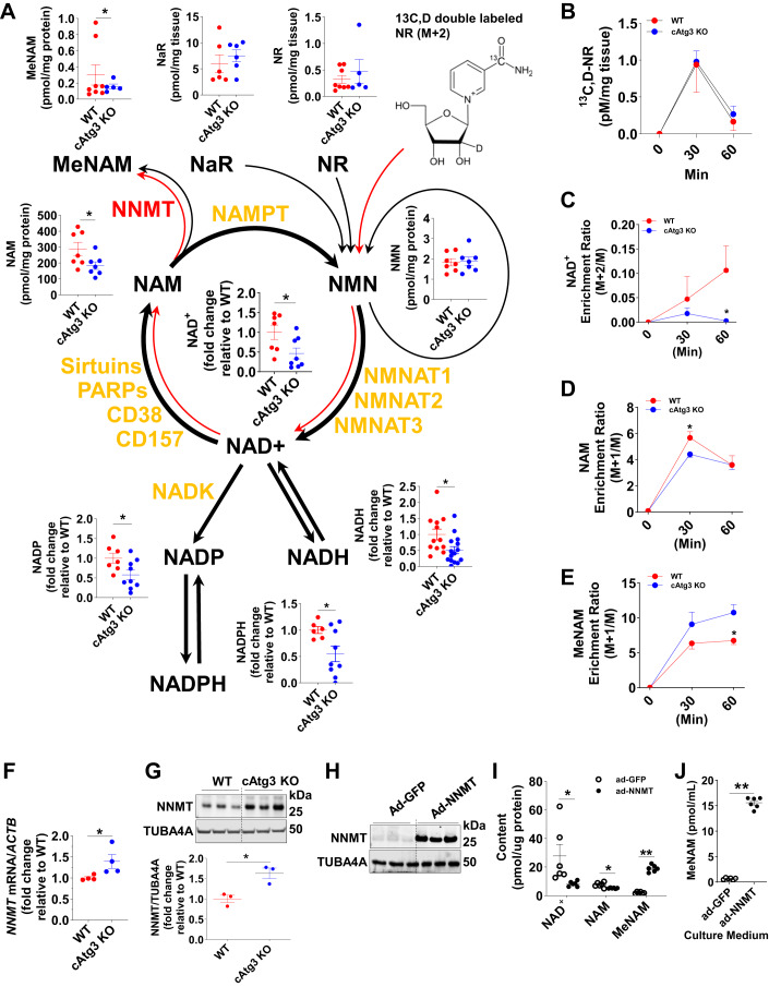
Figure 5. CAtg3-KO mice exhibit altered NAD+ metabolism in hearts.
(A) Schematic of NAD+ catabolic pathways and related metabolites, and NAD+ flux analysis assessed by isotopic nicotinamide riboside (NR) in 6-week-old WT and cAtg3-KO mouse hearts. n = 6–13 per group. Data are mean ± SEM. Unpaired t tests were used to determine statistical significance between two groups. *P < 0.05. (B) Double-labeled NR content in WT and cAtg3-KO mouse hearts at 0, 30 min, and 60 min after isotope-labeled NR administration, n = 4 per group. Data are mean ± SEM. (C–E) NAD+, NAM, and MeNAM enrichment in WT and Atg3 KO hearts at 0 min, 30 min, and 60 min after isotope-labeled NR administration, n = 4 per group. Data are mean ± SEM. Unpaired t tests were used to determine statistical significance between two groups at corresponding time points. *P < 0.05. (F) NNMT mRNA levels in WT and cAtg3-KO mouse hearts at 10 weeks of age, n = 4 per group. Data are mean ± SEM. An unpaired t test was used to determine statistical significance between two groups. *P < 0.05. (G) NNMT and TUBA4A (alpha-tubulin) protein levels in WT and cAtg3-KO mouse hearts at 10 weeks of age, n = 3 per group. Data are mean ± SEM. An unpaired t test was used to determine statistical significance between two groups. *P < 0.05. (H) NNMT and TUBA4A protein expression in H9c2 cells transfected with an adenovirus expressing GFP or NNMT. Representative blots are shown. n = 3 per group. (I) Cellular NAD+, NAM, and MeNAM levels in H9c2 cells transfected with an adenovirus expressing GFP or NNMT, n = 6 per group. Unpaired t tests were used to determine statistical significance between two groups. Data are mean ± SEM, **P < 0.01, *P < 0.05. (J) MeNAM levels in cultured medium from H9c2 cells transfected with adenovirus expressing GFP or NNMT. n = 6 per group. Data are mean ± SEM. An unpaired t test was used to determine statistical significance between two groups. **P < 0.01. Data information: The data in (H–J) were obtained on H9c2 cells. This cell type is derived from embryonic BD1X rat heart tissue. Therefore, H9c2 cells are neither adult cardiomyocytes nor neonatal rat cardiomyocytes. Source data are available online for this figure.

