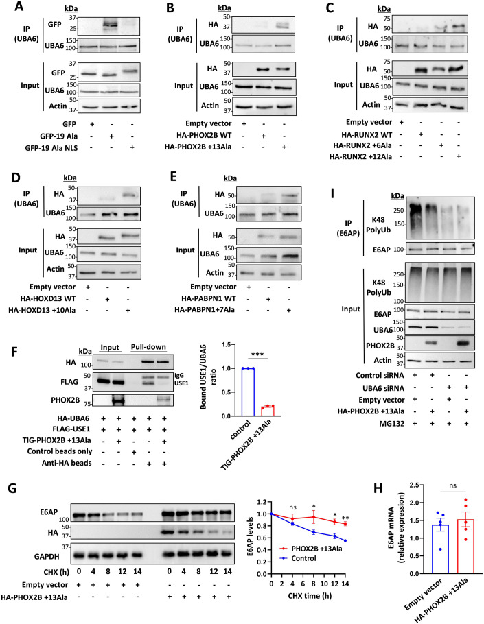
Figure 3. Polyalanine-expanded disease proteins interact with UBA6 and inhibit E6AP degradation.
(A–E) HEK293T cells were transfected with the indicated constructs and immunoprecipitated for endogenous UBA6. (A) Empty GFP, and GFP-polyAla with or without a nuclear localization sequence (NLS). (B) WT and mutant PHOX2B (+13Ala). (C) WT and mutant RUNX2 (+6Ala and +12Ala). (D) WT and mutant HOXD13 (+10Ala). (E) WT and mutant PABPN1 (+7 Ala). (F) Cell lysates of HEK293T cells expressing HA-UBA6 and FLAG-USE1 were incubated with recombinant TIG-mutant PHOX2B. Anti-HA beads were used to pulldown UBA6 (beads only were used as control). The bound USE1/UBA6 ratio is shown. n = 3 biological replicates. (G) Cells were transfected with mutant PHOX2B or empty vector. The cells were treated with cycloheximide (CHX) at the indicated times, and were analyzed for E6AP levels. Results are normalized to t = 0. n = 3 biological replicates. (H) Quantification of E6AP mRNA in the cells transfected with empty vector or mutant PHOX2B. n = 5 biological replicates. (I) Control and UBA6-depleted HEK293T cells were transfected with mutant PHOX2B or empty vector and incubated for the last 6 h with the proteasome inhibitor MG132 (10 μM). Endogenous E6AP was immunoprecipitated from cell lysates for ubiquitination analysis. Data information: Data points in (F–H) represent mean ± s.e.m. P values were calculated by paired 2-tailed t test (F), Two-way ANOVA Sidak’s test (G) and unpaired 2-tailed t test (H). *P < 0.05, **P < 0.01, ***P <0.001, ns non-significant. Source data are available online for this figure.

