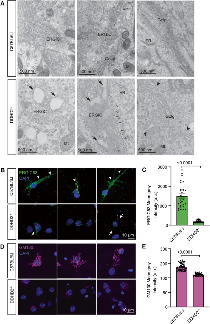
Figure 6. DDHD2 knockout perturbs the secretory pathway.
(A) Selection of representative electron micrographs of the secretory pathway in cultured E16 hippocampal neurons from C57BL/6J and DDHD2−/− mice at DIV21. Images show enlarged Golgi apparatus lumen (arrowheads) and dilated tubulo-vesicular ERGIC (arrows), as well as an accumulation of budding vesicles (open arrowheads) in the ERGIC of DDHD2−/− neurons compared to C57BL/6J neurons. Mitochondria (Mt) and endoplasmic reticulum (ER) are indicated for reference. (B) A selection of representative maximum projections of cultured E16 hippocampal neurons from C57BL/6J and DDHD2−/− mice at DIV21 immunostained against endogenous ERGIC53 (green) and stained with DAPI (blue). Arrows point at ERGIC53 distributed from the perinuclear space to the somatodendritic area. (C) Quantification of ERGIC53 mean grey intensity in C57BL/6J and DDHD2−/− neurons. (D) A selection of representative maximum projection of cultured E16 hippocampal neurons from C57BL/6J and DDHD2−/− mice at DIV21 immunostained against endogenous GM130 (magenta) and stained with DAPI (blue). (E) Quantification of GM130 mean grey intensity in C57BL/6J and DDHD2−/− neurons. Data information: In (A), statistical testing of non-normally distributed data was done using Mann–Whitney U test. n = 30–53 regions of interest (ROIs) from 3 independent experiments, In (C, E), the significance of the difference between each group as determined by unpaired t-test is indicated as <0.0001, and the error bars represent the cumulative standard error of the mean (SEM) for all groups and parameters. n = 30–55 cells from 3 independent experiments.

