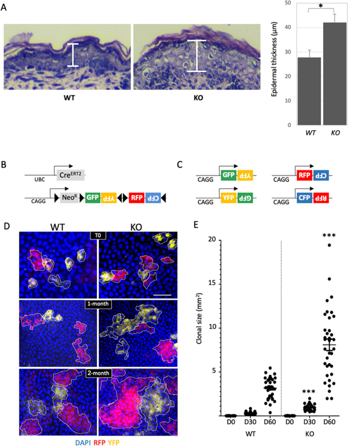
Fig. 1. miR-184-deficient mice display epidermal hyperplasia and enhanced stem cell dynamics.
A Paraffin sections of back skin of newborn mice of the miR-184-wild type (WT) and knockout (KO) mice were used for hematoxylin and eosin (H&E) staining. Quantification is presented on the right panel. B Schematic illustration of the double transgenic lineage tracing system. The Cre driver is controlled by a UBC promoter and the Brainbow2.1 cassette contains a ubiquitously expressed CAG promoter, LoxP (black triangle)-flanked NeoR-roadblock and a head-to-tail LoxP-flanked dimers of green (GFP), yellow (YFP), and red (RFP) and cyan (CFP) fluorescent protein coding genes. C Example of potential rearrangement of the Brainbow2.1 cassette following tamoxifen induced Cre-recombination. D Two-month old mice of the indicated genotypes were treated with tamoxifen for 3–4 days, mice were sacrificed at the indicated day post last treatment, and the basal layer of the tail epidermis was imaged by confocal microscope. E Colonies were identified (dashed line) and their size was defined by imageJ analysis as detailed in Methods. Data represents average and standard deviation from 3 biological replicates. Scale bars were 20 μm. Statistical significance was assessed by t test (*p < 0.05; ***p < 0.001).

