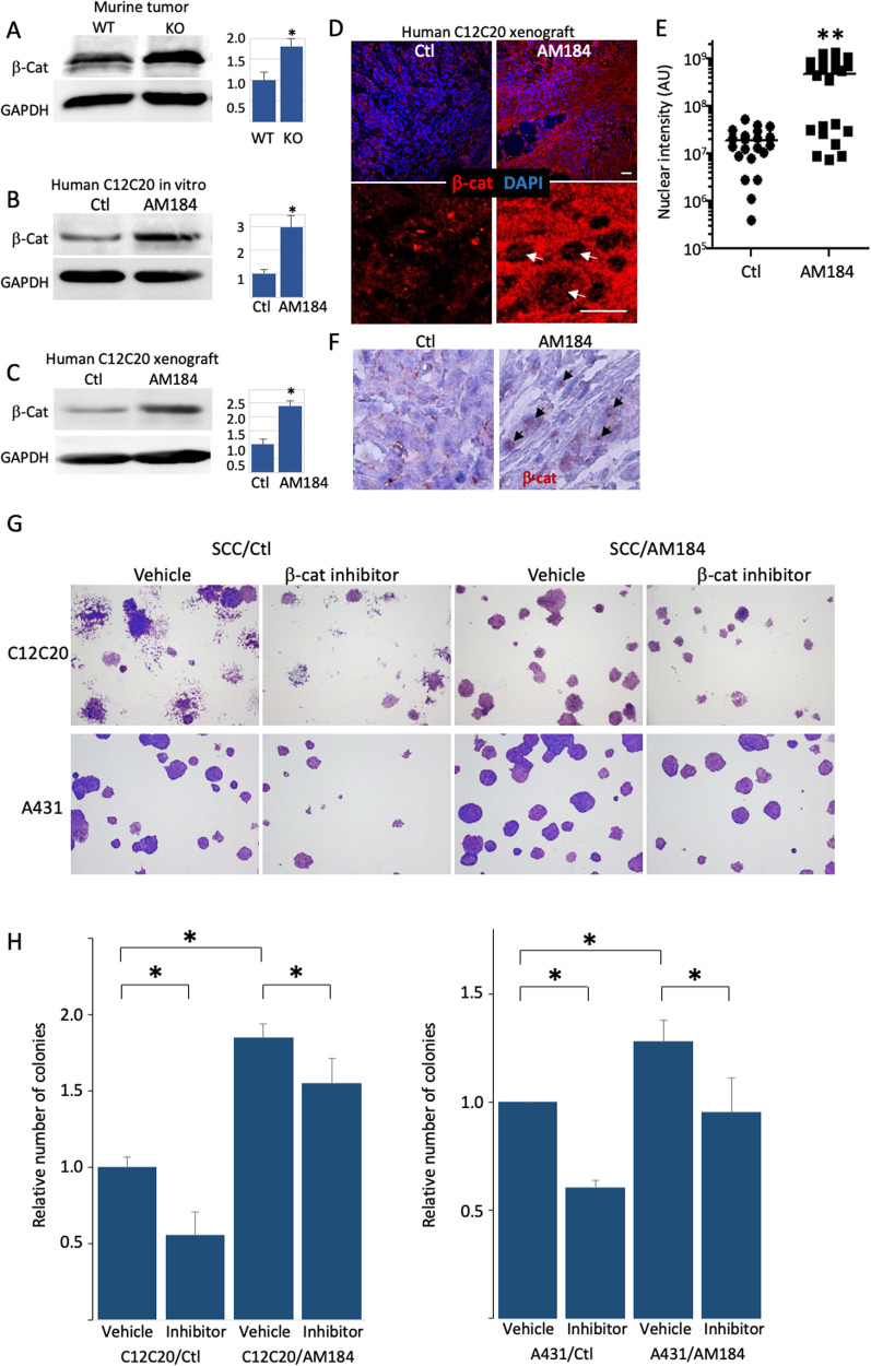
Fig. 5. miR-184-repression of Wnt/β-catenin contributes to SCC phenotype.
A–C The indicated samples of murine wild type (WT) or knockout (KO) tumors or human squamous cell carcinoma C12C20 line that were infected with control (Ctl) or anti-miR-184 antagonist (AM184) were lysed and subjected to Western blot analysis of β-Catenin (β-Cat) or GAPDH as a loading control. Quantified data is shown on the right. Immunofluorescent (D) and its quantification (E) and immunohistochemistry (F) staining showing increased β-Cat staining in tumors of AM184 infected SCC cells. E Quantification of the nuclear signal (arrows in D) was performed by Image J software in 7 different fields in each of the 3 biological replicates. Each dot represents an average of a single field. G, H Colony formation assay performed using the indicated SCC cell line (A431 or C12C20) that were infected with Ctl or AM184 and treated with vehicle or with the β-Catenin/TCF4 inhibitor PKF118-310 as detailed in Methods. Representative image shown in (G) and quantification is shown in (H). Data in represents 3 biological replicates. Scale bars were 20 μm. Statistical significance was assessed by t test (*p < 0.05).

