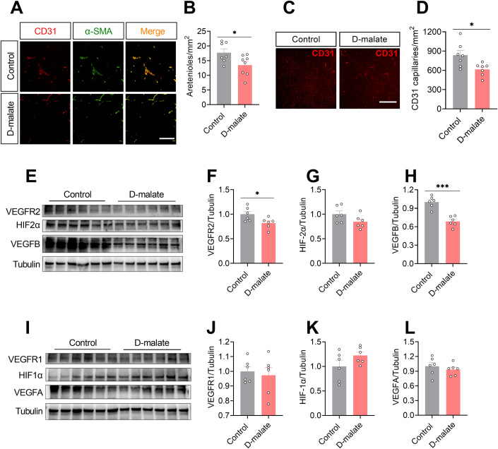
Figure 4. d-malate reduced vascularization and protein expression of VEGFR2 and VEGFB in skeletal muscle.
(A, B) Representative images and co-staining of α-SMA (green) and CD31 (red) (A) and statistical analysis (B) of gastrocnemius in in C57BL/6 male mice after 10 weeks d-malate treatment (n = 7–8 for each group), Scale bars, 200 μm. (C, D) Representative images of CD31 (C) and statistical analysis (D) of gastrocnemius in C57BL/6 male mice after 10 weeks d-malate treatment (n = 7 for each group), Scale bars, 200 μm. (E–H) The protein expression of VEGFR2, HIF2α and VEGFB in the of gastrocnemius in C57BL/6 male mice after 10 weeks d-malate treatment (n = 6 for each group). (I–L) The protein expression of VEGFR1, HIF1α, and VEGFA of gastrocnemius in C57BL/6 male mice after 10 weeks of d-malate treatment (n = 6 for each group). Data information: t test was used in this figure where error bars represent SEM, and *P < 0.05; ***P < 0.001. Source data are available online for this figure.

