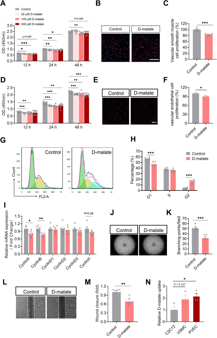
Figure 5. d-malate inhibited the proliferation of vascular smooth muscle cell and vascular endothelial cell and depressed blood vessel formation.
(A) OD value of CCK-8 to detect proliferation activity of vascular smooth muscle cells (n = 6–7 for each group). (B, C) EdU immunofluorescence images (B) and statistics (C) of proliferative activity of vascular smooth muscle cells (n = 5 for each group). Scale bars, 200 μm. (D) OD value of CCK-8 to detect proliferation activity of vascular endothelial cells (n = 9 for each group). (E, F) EdU immunofluorescence images (E) and statistics (F) of proliferative activity of vascular endothelial cells (n = 4 for each group). Scale bars, 200 μm. (G, H) Representative image (G) and statistical graph (H) of vascular endothelial cell cycle by flow cytometry (n = 6 for each group). x axis of G means DNA content. (I) The mRNA expression of cyclin family in vascular endothelial cells (n = 6 for each group). (J, K) Representative images (J) and statistics (K) of vascular endothelial cell tube formation test (n = 6 for each group). (L, M) Representative images (L) and statistics (M) of vascular endothelial cell Scratch test (n = 6 for each group). (N) The relative d-malate uptake in C2C12, vascular smooth muscle cell and vascular endothelial cell after 24 h treatment with 100 μM d-malate (n = 3 for C2C12 and VSMC culture medium, n = 4 for PVEC culture medium). Data information: t test was used in this figure where error bars represent SEM, and *P < 0.05; **P < 0.01; ***P < 0.001. Source data are available online for this figure.

