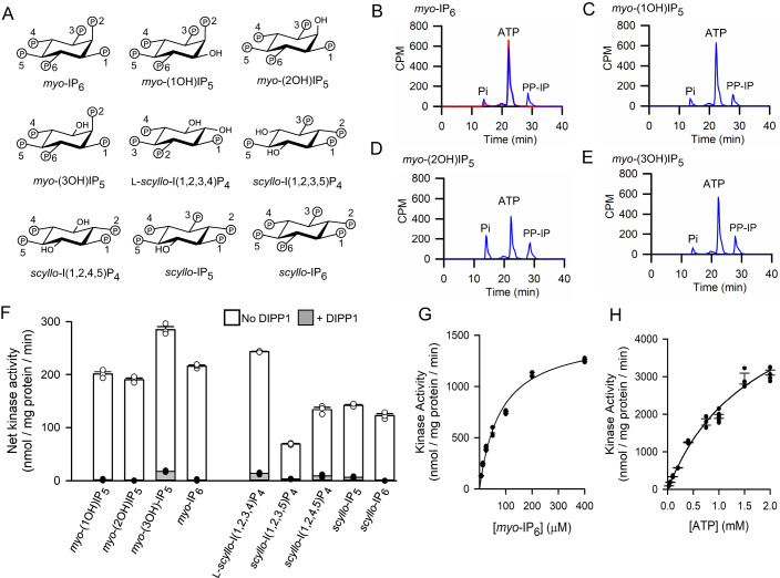
Figure 1. Kinase activities of TvIPK in vitro.
(A) Chemical structures of inositol phosphates incubated with TvIPK in the experiments described in this figure; phosphate numbering follows published guidelines (Murthy, 2006; Pramanik et al, 2020). See Appendix Fig. S1 for structures of other inositol phosphates used in this study. (B–E) Representative (1 of 3 independent experiments) HPLC data following incubations containing [γ-33P]-ATP, either with TvIPK (blue traces) or without enzyme (red trace), plus 50 µM of (B) myo-IP6 or (C) myo-(1OH)IP5 or (D) myo-(2OH)IP5 or (E) myo-(3OH)IP5. (F) PP-IP product accumulation from assays performed either in the absence (open bars) or presence of HsDIPP1 (closed bars) upon (i.e., apparent kinase activity) after incubation of TvIPK with 1 mM ATP and 200 µM of the indicated myo- or scyllo-IPs; (G) Substrate saturation plot for TvIPK incubated with 1 mM ATP and the indicated concentrations of myo-[3H]IP6; activities were analyzed by HPLC. (H) Substrate saturation plot for TvIPK incubated with 2 mM myo-IP6 and the indicated concentrations of [γ-33P]-ATP; activities were analyzed by HPLC. (F–H) show each data point obtained from either 3 or 6 independent experiments (some data points are superimposed); standard errors are also indicated by vertical bars. Source data are available online for this figure.

