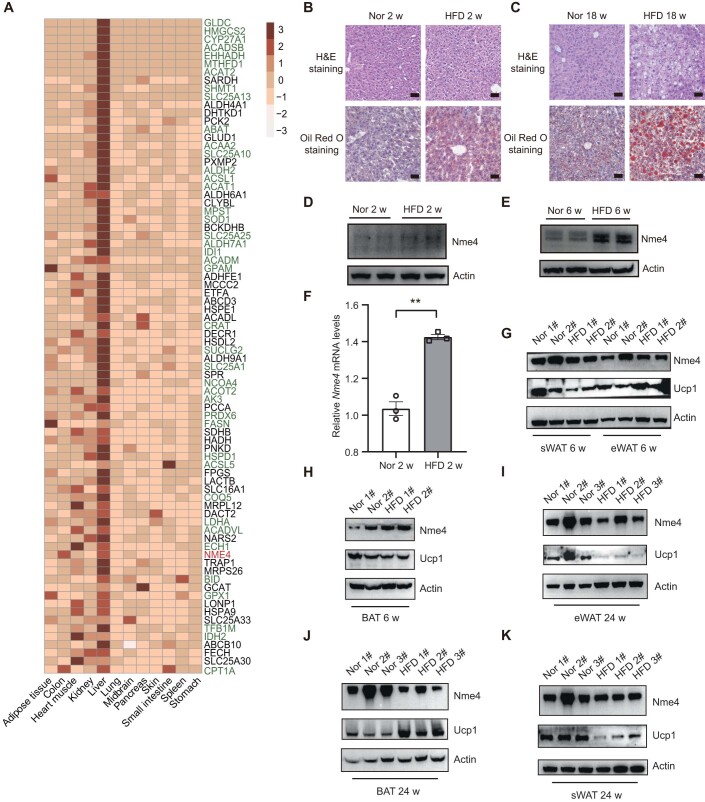
Figure EV1. NME4 is upregulated in fatty liver, and its level is correlated with NAFLD progression.
(A) Heatmap showing the tissue specificity of 80 mitochondrial proteins that were highly expressed in liver tissue. (B, C) H&E staining and Oil Red O staining of liver sections of the mice fed a high-fat diet (HFD) for the indicated times. Scale bars, 50 μm. (D, E) Western blots probed with antibodies against NME4 and Actin in the livers of mice fed a normal diet or a high-fat diet (HFD) for the 2 weeks and 6 weeks. (F) Relative mRNA levels of NME4 in the livers of the mice fed a normal diet or HFD for 2 weeks was measured by RT‒qPCR. Biological replicates, n = 3. (G, H) Western blots probed with antibodies against NME4, UCP1 and Actin in the subcutaneous white adipose tissue (sWAT), epididymal white adipose tissue (eWAT) and brown adipose tissue (BAT) of mice fed a normal diet or a high-fat diet (HFD) for the 6 weeks. (I–K) Western blots probed with antibodies against NME4, UCP1 and Actin in the sWAT, eWAT, BAT and liver of mice fed a normal diet or a high-fat diet (HFD) for the 24 weeks. Data information: (F) data are presented as mean ± SEM. **P values ≤ 0.01 (Student’s t test, unpaired). w, week; HFD high-fat diet, sWAT subcutaneous white adipose tissue, eWAT epididymal white adipose tissue, BAT brown adipose tissue.

